Joshua M. Kruger, MD, PhD | Neuro-Ophthalmology Service, Hadassah Medical Center, Kiryat Hadassah, Jerusalem, Israel Dean M. Cestari, MD | Neuro-Ophthalmology Service, Massachusetts Eye and Ear Infirmary, Harvard Medical School, Boston, Massachusetts Mary Beth Cunnane, MD | Department of Radiology, Massachusetts Eye and Ear Infirmary, Harvard Medical School, Boston, Massachusetts
Neuroimaging is an important tool in ophthalmology, but many ophthalmologists are uncomfortable evaluating actual scans. Unfortunately, exclusive reliance on a radiologist’s report can lead to diagnostic and management errors. We outline a methodology for equipping ophthalmologists with the skills necessary to read neuroimaging studies with respect to specific clinical questions.
Introduction
Neuro-ophthalmic diseases can be divided broadly into disorders that cause afferent or efferent dysfunction. On the afferent side, patients can experience vision loss due to disease affecting the optic nerve, chiasm, optic tract, thalamus, optic radiations, or cerebral cortex. On the efferent side, patients can develop ocular motility abnormalities resulting from lesions anywhere from the cortex to the nerves that supply the extraocular muscles or even from the muscles themselves. Advances in imaging now allow clinicians to identify the locus and often the nature of the lesions responsible for afferent or efferent dysfunction. It is critical for the general ophthalmologist to know which neuro-imaging study to order for each clinical scenario—an issue that we have addressed previously.(1) It is also important for the ophthalmologist to review the study rather than simply to rely on the official radiology report. Oversights can be made by the radiologist. This may occur because the radiologist has limited experience in orbital/neuro-imaging or because they lack the clinical information necessary to identify the specific region requiring attention. We believe that it is possible to train ophthalmologists to be able to read neuro-imaging studies with approaches focused on specific clinical questions. We do not intend to imply that expert radiological evaluation is unnecessary; rather, we hope to provide ophthalmologists with tools to deepen comprehension of basic neuroimaging techniques to permit more informed diagnoses and treatment strategies. The present review aims to provide focused systematic approaches for reviewing various scans and identifying the most common pathologies. The protocols provided in this manuscript require the clinician to be familiar with the various forms of magnetic resonance imaging (MRI; eg, T1, T2, FLAIR). Each sequence highlights various features of neuroanatomy (Table 1; also see Figure 1).
Suspected Optic Neuropathy
Patients with decreased visual acuity, dyschromatopsia, a relative afferent pupillary defect, and a visual field defect should be presumed to have an optic neuropathy. If the patient is >50 years of age, and there is associated optic nerve swelling, then a diagnosis of anterior ischemic optic neuropathy can be presumed, and there is no indication for neuroimaging, provided that the swelling resolves within 2 months.(2) For all other presentations, an immediate MRI of the orbits with contrast is indicated. When optic neuritis is suspected (eg, associated pain with eye movement), then an MRI of the brain with contrast is additionally desirable to assess for radiological evidence of multiple sclerosis.(3,4)
Review of the MRI scan should begin with the coronal short inversion recovery (STIR) or fat-saturated T2-weighted sequences immediately posterior to the globes. The optic nerve can be found at the center of an imaginary ring formed by the extraocular muscles. It is surrounded by a bright cuff of cerebrospinal fluid (CSF). The nerve should be isointense to the subcortical white matter. The clinician should follow the course of the optic nerves all the way to the chiasm, and if possible, into the optic tracts, to ensure that there is symmetry, and isointensity to the subcortical white matter throughout the course (Video 1). The finding of increased brightness (relative to the subcortical white matter) is a nonspecific finding suggestive of injury to the affected segment. The timing cannot be determined, because both acute and chronic injury will cause T2 hyperintensity. Proceed to the coronal T1 post-contrast sequence, preferably with fat saturation. Once again, the optic nerves, chiasm, and tracts should be isointense to the subcortical white matter. The presence of increased brightness (referred to as “enhancement” on a postcontrast image) indicates breakdown of the blood-brain barrier, which may be due to inflammation or tumor. If the nerve does not appear enlarged, then the cause is more likely inflammatory. If optic neuritis is suspected, then one must assess for intracranial evidence of multiple sclerosis, specifically T2 hyperintensities, known as “white spots,” in the brain. Scroll through the axial fluid attenuated inversion recovery (FLAIR) sequence, with particular attention to the periventricular areas. The brainstem is best viewed with T2-weighted imaging. The sagittal FLAIR sequence should also be reviewed to assess for the presence of T2 hyperintense lesions radiating from the corpus callosum, known as Dawson’s fingers (see end of Video 8).
Bitemporal Hemianopsia
A chiasmal disorder is typified by a bitemporal hemianopsia, due to the compression of the crossing fibers from the nasal aspect of each retina. The most common lesion causing a chiasmopathy is a pituitary adenoma,(5) which is typically benign, but the differential diagnosis includes disorders such as an aneurysm(6) and pituitary apoplexy.(7) The chiasm can be imaged through either an MRI orbit sequence or an MRI brain scan. Many institutions also offer the option of a specific pituitary scanning protocol.
Review of the scan (Video 2) should begin with a sagittal image, preferably T1. The pituitary gland can be identified as an area of soft tissue within the bony sella, which lies along the superior-posterior aspect of the dark sphenoid sinus. The posterior segment of the pituitary gland is particularly bright, likely due to the presence of lipid in the posterior pituicytes.(8) The normal height of the pituitary gland is approximately 5 mm,(9) but it can be as much as 9 mm.(10) Immediately superior to the pituitary gland is the suprasellar cistern, which will appear dark on T1 coronal imaging. Above the suprasellar cistern is the optic chiasm, which appears as a bright, linear structure oriented at approximately 45 degrees on T1 imaging. The presence of a complete suprasellar cistern indicates that the pituitary cannot be exerting a mass effect on the chiasm.
Next, review the coronal T1 sequence. If it is postcontrast, then the pituitary will be diffusely bright because it is a highly vascularized structure. A T-shaped structure extends upward from the center of the gland—the vertical component is the pituitary stalk, and the horizontal component is the optic chiasm. Once again ensure that CSF clearly separates the chiasm from the pituitary gland. Video 3 demonstrates findings for a pituitary adenoma. Note that the described approach has not included review of the axial scan because the chiasm is poorly visualized in that plane.
Homonymous Hemianopsia
A visual field defect that occurs right or left of the vertical midline in both eyes is referred to as “homonymous” and is associated with a retrochiasmal disorder. These cases are best imaged through an MRI of the brain. If the visual field loss has an acute onset in an elderly patient, then the mechanism likely is due to a stroke, and it is reasonable to forgo contrast because this is not useful for demonstrating ischemia. The critical sequence is diffusion weighted imaging (DWI), which is often performed as part of a standard MRI but should be explicitly requested in these circumstances. If it is a case of homonymous visual defect in a younger patient, a mass or demyelinating lesion is more likely and contrast should be included.
The visual pathway extending from the origin of the optic tracts to the occipital cortex must be inspected. The optic tracts can be identified in both axial and coronal images. They extend posteriorly from the optic chiasm and course around the midbrain to the lateral geniculate body of the thalamus. The optic radiations run from the lateral geniculate body to the occipital cortex. En route, they spread diffusely within the white matter of the temporal and parietal lobes. The optic radiations are best examined with axial images. The DWI, FLAIR, and T1 pre- and postcontrast images should be inspected for hyperintense lesions in the areas described. A hyperintense lesion in DWI can represent either cytotoxic edema (ie, stroke) or vasogenic edema. If the edema is cytotoxic, then the same region will appear hypointense on the corresponding apparent diffusion coefficient (ADC) images. An area that is hyperintense on DWI, hypointense on ADC, and hyperintense on T2-weighted imaging (eg, FLAIR) most likely represents a subacute infarction (Video 4). The additional presence of hyperintensity on noncontrast T1-weighted images is concerning for an intracerebral hemorrhage. The presence of hyperintensity specifically on the postcontrast images only (and not present on the T1 precontrast images) is suggestive of a neoplastic lesion, vascular lesion, inflammation, or infection.
Oculomotor Nerve Palsy
The third cranial nerve innervates the superior rectus, inferior rectus, medial rectus, and inferior oblique muscles as well as the levator palpebrae superioris. It also carries the parasympathetic pupillary constrictor fibers. In the most extreme form of an oculomotor nerve palsy, there is complete ptosis, a dilated pupil, and the globe will assume a down-and-out position. It is critical, however, to keep in mind that many presentations of an oculomotor nerve palsy can affect only some of these muscles, often in a very mild way. The index of suspicion must be very high in any case of diplopia with a vertical component. Because the oculomotor nerve enters the subarachnoid space, it comes into close proximity with the posterior communicating artery. An aneurysm of this artery may compress the oculomotor nerve, resulting in a palsy. Rupture of an aneurysm is a life-threatening event.(11) Therefore, any oculomotor nerve palsy must be presumed to be caused by a posterior communicating aneurysm until proven otherwise. Computed tomography angiography (CTA) can be just as sensitive as magnetic resonance angiography (MRA) in detecting these aneurysms(12) and delaying imaging to obtain an MRA versus a CTA is not justified. Communication with the neuroradiologist is best to establish the preferred modality for diagnosis of these aneurysms.
Aneurysms are most commonly located at sites of vessel bifurcation; therefore, examination of the angiogram should be focused on the circle of Willis (Video 5). The routes of all major vessels of the anterior and posterior circulation should be inspected, with particular attention to the posterior communicating artery (PCOM). Any outpouching of contrast that extends off a vessel but does not extend into another known vessel is suspicious for an aneurysm. Follow the route of each internal carotid artery (ICA) looking carefully at the take-off for the PCOM, a branch of the supraclinoid internal carotid artery. Continue to follow the ICA as it bifurcates into the anterior and middle cerebral arteries. Follow the course of each middle cerebral artery through the bifurcations into the superior and inferior segments. Follow the course of each anterior cerebral artery, attempting to identify the anterior communicating artery, a tiny vessel which connects the right and left anterior cerebral artery (ACA). Proceed to the posterior circulation, beginning with the vertebral arteries at the level of the foramen magnum. As one scrolls caudally, one should note the origin of the posterior inferior cerebellar arteries (PICAs) off each vertebral artery. The vertebral arteries then merge into the basilar artery. Areas of contrast will then extend off the basilar artery toward the cerebellum, corresponding to the anterior inferior cerebellar arteries (AICA) and the superior cerebellar arteries, respectively. Finally, the basilar artery will bifurcate into left and right posterior cerebral arteries. Volume-rendering reconstruction technologies, such as coronal maximum intensity projection (MIP) sequences or 3-D reconstructions may allow for better appreciation of any aneurysms. Keep in mind that patients may have multiple aneurysms, and that a complete search of the circle of Willis should be performed even in patients with aneurysms large enough to be appreciated on the initial cursory review.
If there is radiological evidence of an aneurysm in the setting of a headache, then the computed tomography (CT) scan should be reviewed to rule out the presence of a subarachnoid hemorrhage (ie, a ruptured aneurysm). Particular attention should be paid for any hyperdensity in the basilar cisterns and ventricles.
Review of an MRI scan for an oculomotor nerve palsy begins with the axial T2-weighted sequence at the level of the midbrain (Video 6). The third cranial nerve is located posterior to the red nucleus, just lateral to the sagittal midline. Ensure that there is no focal hyperintensity in the area. This same region should also be viewed through the DWI sequence to rule out any focus of hyperintensity, which might indicate an acute infarction. The third nerve exits the brainstem anteriorly, just lateral to the interpeduncular cistern, and travels anteriorly toward the cavernous sinus. This segment is best viewed with heavily T2-weighted images (eg, FIESTA, CISS, 3D DRIVE). Note that the third cranial nerve runs parallel to the posterior communicating artery. Coronal postcontrast images throughout the cavernous sinus should then be viewed. The oculomotor nerve is located at the superior lateral aspect of the cavernous sinus. It should be isointense to the subcortical white matter. Symmetry should be confirmed.
Trochlear Nerve Palsy
In the case of an isolated trochlear nerve palsy (or abducens nerve palsy) in a patient >50 years of age who is diabetic or vasculopathic, the most likely etiology is microvascular ischemia, in which case neuroimaging will be unrevealing. In these circumstances, neuroimaging can be deferred as long as there is no worsening of the strabismus,(13) and the strabismus resolves within 90 days. If either of these conditions cannot be met, then an MRI of the brain should be performed with contrast. In the case of a young, nondiabetic patient, a structural cause must be presumed, and an MRI of the brain with contrast should be performed at the soonest opportunity.
Review of the scan begins with the axial FLAIR at the level of the midbrain (Video 7). Located at the posterior aspect of the midbrain is the cerebral aqueduct, which appears as a dark flow void, surrounded by the bright ring of the periaqueductal gray. The trochlear nerve nucleus lies immediately anterior to the periaqueductal gray. Ensure that there is no focal brightness in the area, or hyperintensity on DWI. The trochlear nerve exits the brainstem posteriorly and then travels anteriorly toward the cavernous sinus through the basilar cistern. As stated previously, the cisternal segments of the cranial nerves are best studied with heavily T2-weighted images. While the size of the trochlear nerve is typically too small to be visualized, it is important to rule out the presence of any mass lesions in the basilar cistern, which would be a presumed etiology for a trochlear nerve palsy. The basilar cistern should also be viewed in axial postcontrast images to rule out an enhancing mass or leptomeningeal enhancement. Coronal postcontrast images throughout the cavernous sinus should then be reviewed. Again, the trochlear nerve is typically too small to be seen but sits immediately inferior to the oculomotor nerve, adjacent to the lateral wall of the sinus. This region should be reviewed to rule out the presence of any abnormal mass. If a chronic trochlear nerve palsy is suspected, ensure that the sizes of the superior oblique muscles are symmetric by viewing them in a coronal sequence within the orbits.
Abducens Nerve Palsy
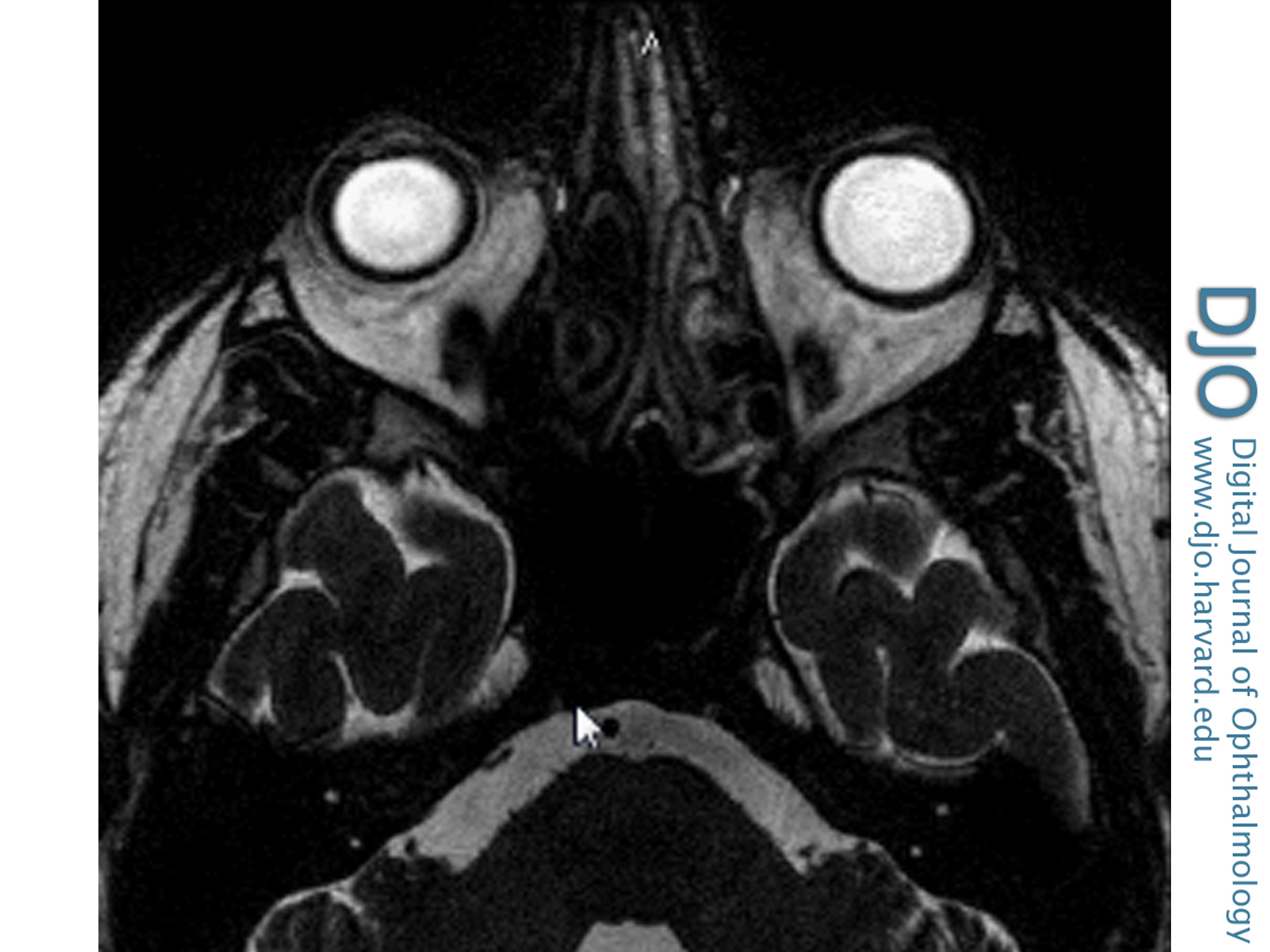
The abducens nerve lies at the dorsal aspect of the caudal pons. It lies immediately ventral to the facial colliculli, which are protrusions of the dorsal pons into the fourth ventricle. Each colliculus is formed by the motor fibers of the facial nerve wrapping around the nucleus of the abducens nerve. This area should be assessed for any hyperintensity in the axial FLAIR, DWI, and postcontrast T1 sequences (Video 8). The abducens nerve exits the brainstem ventrally at the junction of the pons and medulla. It then ascends toward the cavernous sinus through the pontine cistern. Within the cisternal segment, the nerve can be visualized with heavily T2-weighted images, appearing as a linear hypointensity nearly parallel to the sagittal plane. Assess for the presence of any abnormal soft tissue in the heavily T2-weighted and T1 postcontrast images (Video 9). The abducens nerve transitions from the pontine cistern into the cavernous sinus via Dorello’s canal, a groove in the petrous bone. It can be visualized on the axial T2 images as a linear hyperintensity extending from the pontine cistern into the hypointense petrous bone (Figure 2). Coronal postcontrast images throughout the cavernous sinus should then be reviewed. While the oculomotor and trochlear nerves are adjacent to the lateral wall of the cavernous sinus, the abducens nerve is more central, situated inferior-lateral to the internal carotid artery. The caliber is typically too small to be visualized, but the observer should be alert for a T2 hyperintense or enhancing lesion inferolateral to the carotid artery, which would be likely to produce an abducens nerve palsy.
Skew Deviation
A skew deviation is an acquired vertical strabismus caused by a supranuclear lesion. There is defective input to the oculomotor and trochlear nerves, which control the vertical position of the globe. Relevant lesions can occur in the peripheral vestibular system, the brainstem, the cerebellum or the thalamus.(14) These structures can be assessed best on axial T2-weighted imaging (Video 10).
Thyroid Eye Disease
The most common etiology of an orbitopathy causing diplopia is Thyroid eye disease. It is perhaps the best exception to the “rules” that MRI is superior to CT and that it is always preferable to use contrast. CT is the preferred modality to view the extraocular muscles, but iodinated contrast has the potential for worsening the systemic manifestations of thyroid disease and compromising treatment for thyroid malignancies.(15) Thus, order CT imaging without contrast.
Begin review of the scan with an axial sequence at the level of the horizontal rectus muscles (Video 11). The medial rectus muscle should be slightly thicker than the lateral rectus muscle. Most of the globe should be posterior to an imaginary line drawn between the most anterior portions of the medial and lateral orbital walls. There should be no signs of fat proliferation. In the coronal images, the size of the rectus muscles should be assessed immediately posterior to the globe. The sizes of the superior muscle complex, the medial rectus, and the inferior rectus should be roughly equal and symmetric between the orbits. The lateral rectus muscle should be thinner. Move posteriorly to the orbital apex where the four rectus muscles begin to coalesce into the annulus of Zinn. Ensure that there is a rim of fat that separates the optic nerve from the rectus muscles (to rule out compressive optic neuropathy). Video 12 demonstrates a case of thyroid eye disease.
Papilledema
Papilledema is defined as optic nerve swelling that is secondary to increased intracranial pressure. Neuroimaging is mandated prior to performing a lumbar puncture due to the possibility of herniation with opening of the intracranial space, and a CT scan is adequate in urgent situations.(16) The preferred modality is an MRI and magnetic resonance venography (MRV) of the brain with contrast, which has the highest likelihood of detecting a structural lesion. When reviewing the scan, the first objective is to rule out the presence of an intracranial mass, hemorrhage, or hydrocephalus. Examine the midline structures to ensure there is no midline shift. Look at the ventricles to evaluate for ventricular enlargement. Ensure that the sulci extend all the way to the inner calvarium and are not separated from it by a subdural hematoma. Look for displaced structures, swollen regions of the brain, and mass effect. The clinician should then proceed to assess for signs of increased intracranial pressure, which can be seen in idiopathic intracranial hypertension. Begin with the sagittal T1 images and identify the pituitary gland. Increased intracranial pressure causes the CSF in the suprasellar cistern to compress the pituitary gland against the floor of the sella resulting in a “partially empty sella” (Video 13). On the axial T2-weighted images, the anterior aspect of the CSF cuff of the optic nerve should be inspected. In normal individuals, mild flaring can occur (Video 14). Larger amounts of CSF fluid are concerning for increased intracranial pressure (Video 13). The posterior aspect of the globe should have a spherical appearance. It typically becomes “flattened” in idiopathic intracranial hypertension (IIH).
MRV should also be performed in all patients before IIH is presumed, particularly in atypical cases, to rule out the possibility of a sinus venous thrombosis.(17) The course of each sinus should be reviewed in both the raw data and the MIP images. The presence of a filling defect in the lumen of a sinus is known as the empty delta sign and is specific for a thrombus.(18) Stenosis of the transverse sinuses is a common finding in IIH (see Video 15).(19)
Horner Syndrome
Two causes of anisocoria that require urgent neuroimaging are an oculomotor nerve palsy, discussed above, and a Horner syndrome. A Horner syndrome is caused by decreased sympathetic stimulation to the eye, resulting in miosis and ptosis due to disrupted innervation of the pupillary dilators and Muller’s muscle. The sympathetic pathway runs a circuitous course, descending from the hypothalamus to the level of the thoracic spine and then ascending to the orbit.
Until proved otherwise, any case of a Horner syndrome must be presumed to be due to a carotid dissection. Imaging of the carotid artery should be performed using a CTA of the head and neck. The goal is to trace the entire course of each carotid artery, ensuring that the artery retains its full caliber throughout its path. The scan is reviewed with axial sections. Begin at the level of the aortic arch (Video 16). As one scrolls superiorly, the aortic arch transitions into the brachiocephalic (that is, innominate), left common carotid, and left subclavian arteries. Starting on the right, the brachiocephalic divides into the right subclavian artery (posteriorly) and common carotid artery (anteriorly). The common carotid will then bifurcate into the external carotid artery (anteriorly) and the internal carotid artery (posteriorly). The internal carotid artery then ascends through the neck and enters the skull base through the carotid canal, in the petrous bone. It then runs transversely across the bony space within the petrous segment, which it should completely fill. Moving further superiorly, the internal carotid artery passes through the cavernous sinus. The internal carotid artery exits the cavernous sinus and enters the dura roughly at the level of the anterior clinoid. The supraclinoid internal carotid artery then bifurcates into the middle cerebral artery and the anterior cerebral artery. Volume rendering reconstruction technologies, such as MIP, allow for a sagittal view of the internal carotid artery, which is often more readily followed (Video 16). Any tapered narrowing of the internal carotid artery is concerning for the possibility of a dissection. In some cases, MRI may be useful to evaluate for a high signal crescent around the carotid on fat-saturated T1-weighted images. This high signal crescent represents clot in the false lumen of the vessel.
While ruling out a carotid dissection is a high priority in a Horner syndrome, imaging must be performed to scan the pathway of the three orders of neurons involved in supplying sympathetic innervation to the eye. The first-order neuron originates in the hypothalamus. Thus, an MRI of the brain is required to properly image this area. An apical lung tumor (Pancoast tumor) can injure the second-order neuron, and a CT of the chest should be performed to rule out this possibility. Imaging of the third-order neuron is achieved in the previously mentioned imaging of the carotid artery. Therefore, Horner syndrome requires a variety of imaging modalities to rule out these structural causes in the differential diagnosis.
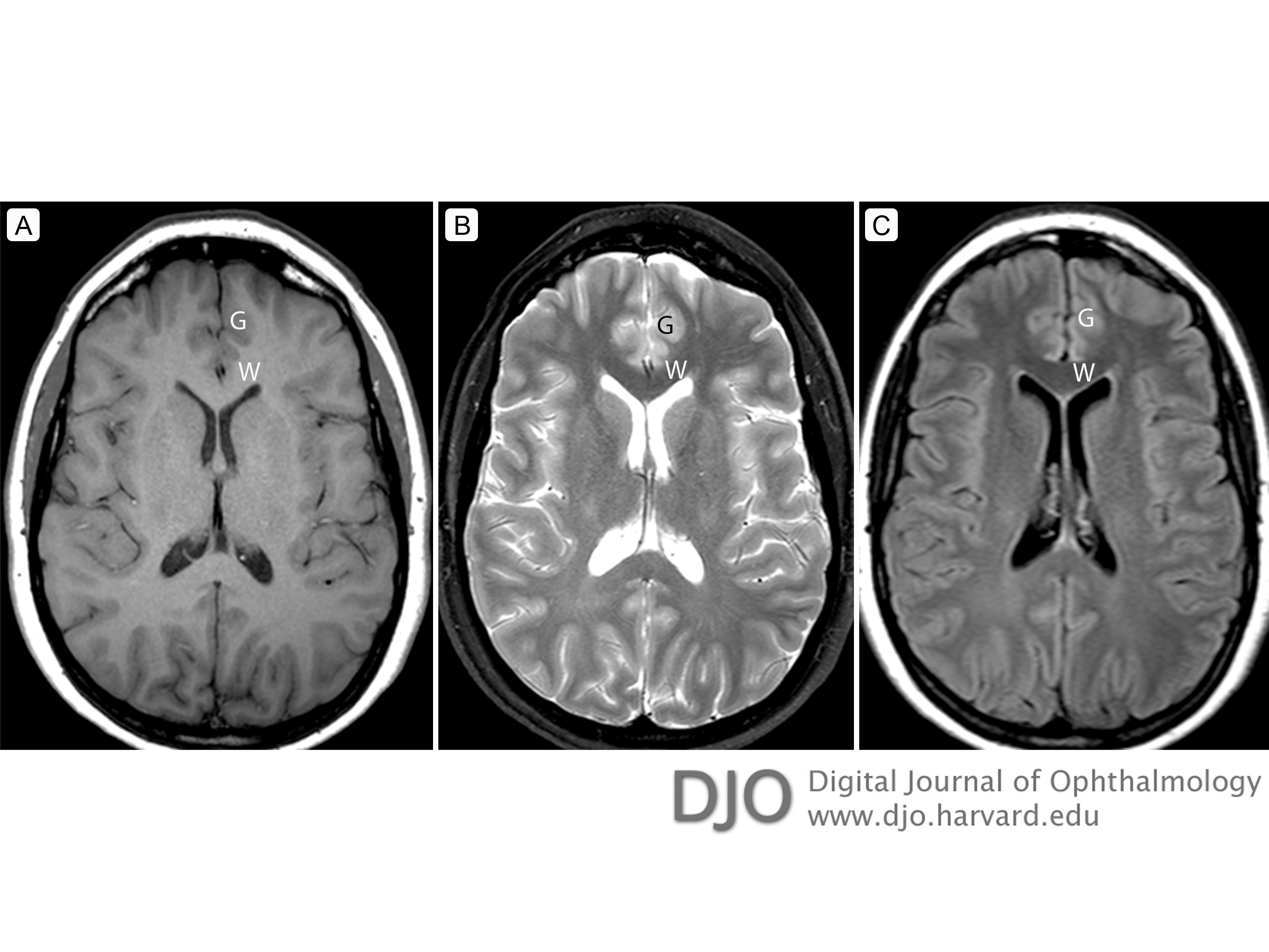
Figure 1
Axial images through the lateral ventricles demonstrate differences in the appearance of T1-weighted, T2-weighted and FLAIR images. In each image, a W is placed over the corpus callosum, a white matter tract, and a G is placed over the cortex of the left frontal lobe. A, On this T1-weighted image, the white matter is hyperintense, and the cerebrospinal fluid (CSF) in the ventricles is hypointense compared to the gray matter. B, On this T2-weighted image, the white matter is hypointense, and the CSF in the ventricles is hyperintense compared to the gray matter. C, A FLAIR image is a T2-weighted image with suppression of the bright CSF signal. On these images, the white matter is hypointense (like a T2-weighted image), but the CSF in the ventricles is hypointense in comparison to the gray matter.
Figure 2
Axial highly T2-weighted scan demonstrates Dorello’s canals as linear hyperintensities extending from the pontine cistern. The right canal is demonstrated with an arrow.
Table 1.
Comparison of MRI sequences
Video 1.
Normal optic nerve.
Video 2.
Sella turcica and pituitary.
Video 3.
Pituitary adenoma.
Video 4.
Occipital lobe infarction.
Video 5.
Computed tomography (CT) angiography for an oculomotor nerve palsy.
Video 6.
Oculomotor nerve.
Video 7.
Trochlear nerve.
Video 8.
Abducens nerve.
Video 9.
Patient with bilateral abducens nerve palsy.
Video 10.
Imaging for cases of skew deviation.
Video 11.
Normal orbit.
Video 12.
Thyroid orbitopathy.
Video 13.
Idiopathic intracranial hypertension – magnetic resonance imaging (MRI).
Video 14.
Idiopathic intracranial hypertension ruled out – MRI.
Video 15.
Idiopathic intracranial hypertension – MR venography.
1. Kruger JM, Lessell S, Cestari DM. Neuro-imaging: a review for the general ophthalmologist. Semin Ophthalmol 2012;27:192-6.
2. Lee AG, Lin DJ, Kaufman M, Golnik KC, Vaphiades MS, Eggenberger E. Atypical features prompting neuroimaging in acute optic neuropathy in adults. Can J Ophthalmol 2000;35:325-30.
3. Polman CH, Reingold SC, Banwell B, et al. Diagnostic criteria for multiple sclerosis: 2010 revisions to the McDonald criteria. Ann Neurol 2011;69:292-302
4. Optic Neuritis Study Group. Multiple sclerosis risk after optic neuritis:final optic neuritis treatment trial follow-up. Arch Neurol 2008;65:727-32.
5. Abboud CF, Laws ER Jr. Diagnosis of pituitary tumors. Endocrinol Metab Clin North Am 1988;17:241-80.
6. Kasner SE, Liu GT, Galetta SL. Neuro-ophthalmologic aspects of aneurysms.Neuroimaging Clin N Am 1997;7:679-92.
7. Nawar RN, AbdelMannan D, Selman WR, Arafah BM. Pituitary tumor apoplexy: a review. J Intensive Care Med 2008;23:75-90.
8. Kucharczyk W, Lenkinski R, Kucharczyk J, Henkelmann RM. The effect of phospholipid vesicles on the NMR relaxation of water: an explanation for the appearance of the neurohypophysis? AJNR Am J Neuroradiol 1990;11:693-700.
9. Tsunoda A, Okuda O, Sato K. MR height of the pituitary gland as a function of age and sex: especially physiological hypertrophy in adolescence and in climacterium. AJNR Am J Neuroradiol 1997;18:551-4.
10. Wolpert SM, Molitch ME, Goldman JA, Wood JB. Size, shape, and appearance of the normal female pituitary gland. AJR Am J Roentgenol 1984;143:377-81.
11. Huige WM, van Vliet AG, Bastiaensen LA. Early symptoms of subarachnoid haemorrhage due to aneurysms of the posterior communicating artery. Doc Ophthalmol 1988;70:251-6.
12. Lee AG, Hayman LA, Brazis PW. The evaluation of isolated third nerve palsy revisited: an update on the evolving role of magnetic resonance, computed tomography, and catheter angiography. Surv Ophthalmol 2002;47:137-57.
13. Chi SL, Bhatti MT. The diagnostic dilemma of neuro-imaging in acute isolated sixth nerve palsy. Curr Opin Ophthalmol 2009;20:423-9.
14. Wong AM. Understanding skew deviation and a new clinical test to differentiate it from trochlear nerve palsy. J AAPOS 2010;14:61-7.
15. van der Molen AJ, Thomsen HS, Morcos SK; Contrast Media Safety Committee, European Society of Urogenital Radiology (ESUR). Effect of iodinated contrastmedia on thyroid function in adults. Eur Radiol 2004;14:902-7.
16. Gower DJ, Baker AL, Bell WO, Ball MR. Contraindications to lumbar puncture as defined by computed cranial tomography. J Neurol Neurosurg Psychiatry 1987;50:1071-4.
17. Lin A, Foroozan R, Danesh-Meyer HV, De Salvo G, Savino PJ, Sergott RC. Occurrence of cerebral venous sinus thrombosis in patients with presumed idiopathic intracranial hypertension. Ophthalmology 2006;113:2281-4.
18. Virapongse C, Cazenave C, Quisling R, Sarwar M, Hunter S. The empty delta sign: frequency and significance in 76 cases of dural sinus thrombosis. Radiology1987;162:779-85.
19. Riggeal BD, Bruce BB, Saindane AM, et al. Clinical course of idiopathic intracranial hypertension with transverse sinus stenosis. Neurology 2013;80:289-95.
 Welcome, please sign in