|
|
 |
|
|
|
|
|
Primary orbital melanoma in association with cellular blue nevus
Digital Journal of Ophthalmology 2014
Volume 20, Number 3
July 14, 2014
DOI: 10.5693/djo.01.2014.02.001
|
Printer Friendly
Download PDF |

Tarek El-Sawy , MD, PhD | Orbital Oncology Ophthalmic Plastic Surgery Program, Department of Plastic Surgery, The University of Texas MD Anderson Cancer Center, Houston, Texas Mathieu F. Bakhoum, PhD | Orbital Oncology & Ophthalmic Plastic Surgery Program, Department of Plastic Surgery, The University of Texas MD Anderson Cancer Center, Houston, Texas Michael Tetzlaff, MD, PhD | Department of Pathology, The University of Texas MD Anderson Cancer Center, Houston, Texas Qasiem J. Nasser, MD | Orbital Oncology Ophthalmic Plastic Surgery Program, Department of Plastic Surgery, The University of Texas MD Anderson Cancer Center, Houston, Texas Victor G. Prieto, MD, PhD | Department of Pathology, The University of Texas MD Anderson Cancer Center, Houston, Texas Doina Ivan, MD | Department of Pathology, The University of Texas MD Anderson Cancer Center, Houston, Texas Matthew C. Sniegowski, MD | Orbital Oncology Ophthalmic Plastic Surgery Program, Department of Plastic Surgery, The University of Texas MD Anderson Cancer Center, Houston, Texas Vivian T. Yin, MD | Orbital Oncology Ophthalmic Plastic Surgery Program, Department of Plastic Surgery, The University of Texas MD Anderson Cancer Center, Houston, Texas Caroline Pan, MD | Department of Ophthalmology, The University of Colorado School of Medicine, Denver, Colorado Vikram Durairaj, MD | Department of Ophthalmology, The University of Colorado School of Medicine, Denver, Colorado Bita Esmaeli, MD | Orbital Oncology & Ophthalmic Plastic Surgery Program, Department of Plastic Surgery, The University of Texas MD Anderson Cancer Center, Houston, Texas
|
|
|
| Abstract | Purpose
To describe 3 cases of primary orbital melanoma associated with either known or subsequently discovered cellular blue nevus.
Methods
The clinical records and surgical specimens of 3 patients who underwent orbital exenteration for primary orbital melanoma and who had a cellular blue nevus diagnosed before or after detection of the melanoma were retrospectively reviewed.
Results
All 3 patients presented with signs and symptoms of an orbital mass. Subsequent biopsy revealed invasive melanoma. One patient had a known history of congenital cellular blue nevus of the eyelid from which the orbital melanoma originated. The other 2 patients had no known history of cutaneous pigmentation or blue nevus. In these 2 patients, the cellular blue nevus was detected on pathologic review of the orbital exenteration specimen (1 patient) or surgical biopsy specimen (1 patient). All 3 patients underwent total body positron emission tomography/computed tomography, and in all 3 results were negative for other sites of disease involvement. In the 2 patients without a previously known nevus a total body skin check was negative for other primary melanoma lesions. All 3 patients underwent orbital exenteration followed by postoperative radiation therapy.
Conclusions
Thorough evaluation of biopsy specimens of “primary” orbital melanoma is warranted to ensure identification of any associated blue nevus because blue nevi are precursor lesions for orbital melanoma, and the presence of a blue nevus would support a primary orbital melanoma rather than a metastatic lesion. Patients with a known blue nevus of the periocular skin and ocular adnexa should be monitored closely for signs of malignant transformation. | | | Introduction | Primary orbital melanoma is exceedingly rare and estimated to account for fewer than 1% of orbital tumors in most large series.(1,2) The vast majority of orbital melanomas are either metastatic or represent extension from skin and conjunctival melanomas.(3) Distinguishing primary orbital melanoma from metastatic orbital melanoma or extension from adjacent structures is essential because the treatment for primary melanoma of the orbit is usually orbital exenteration, whereas exenteration would only be appropriate as a palliative measure in cases of metastasis or extension from adjacent orbital structures.
Blue nevus and cellular blue nevus are benign, intradermal melanocytic tumors that are believed to represent the arrest of neural crest melanocyte precursors within the dermis. They differ in degree of cellularity. The presence of an associated cellular blue nevus on pathologic examination of an orbital melanoma provides evidence supporting a primary orbital melanoma because the cellular blue nevus component is a precursor lesion.(4,5) We report 3 cases of primary orbital melanoma associated with a cellular blue nevus. | | | Materials and Methods | | This study was exempt from institutional review board review because of lack of aggregate data. The clinical records of 3 patients with primary orbital melanoma who had a diagnosis of a cellular blue nevus either before or after detection of the primary orbital melanoma were retrospectively reviewed. Parameters recorded from each patient’s chart included age at the time of diagnosis, sex, results of systemic imaging work-up, results of pathologic examination of the diagnostic biopsy specimen, surgical treatment, results of pathologic examination of the final surgical specimen, duration and dose of postoperative radiotherapy, length of follow-up, and disease status at last appointment. | | | Results | Patient 1
A previously healthy 9-year-old girl with a known history of blue nevus involving her right lower eyelid and cheek since birth (Figure 1) presented to MD Anderson Cancer Center, Houston, with progressive proptosis of the right eye and swelling of the right lower eyelid. A surgical debulking procedure by her local oculoplastic surgeon a few months earlier confirmed the presence of invasive melanoma in the orbit. She was referred to MD Anderson for further management.
On examination at our center, visual acuity was 20/20, ocular motility was full, without diplopia, and visual fields were full. Magnetic resonance imaging (MRI) revealed an orbital mass with direct extension into the pterygopalatine fossa along the inferior orbital fissure (Figure 2) and approaching the superior orbital fissure. Systemic work-up included a total body positron emission tomography (PET), computed tomography (CT) scan, ultrasonography of regional lymph nodes, and chest radiography. All scans were negative for metastatic disease.
The patient underwent an orbital exenteration, partial maxillectomy, partial ethmoidectomy, partial sphenoidectomy, and dissection of the deep orbital apical and skull base extension of the lesion performed by a multidisciplinary surgical team, including Oculoplastics, Head and Neck Surgery, and Neurosurgery. Pathologic examination of the surgical specimen revealed several foci of invasive melanoma associated with an atypical cellular blue nevus (Figure 3). There was extensive cellular blue nevus within the orbit, conjunctiva, and skin. The margins of the resection specimen were negative for melanoma. Immunochemical staining for markers of melanocytic differentiation was performed. HMB-45 was diffusely positive in the tumor cells (not shown). An anti-MART1/Ki-67 cocktail was used to assess the proliferative index of the melanocytes comprising the lesion revealed the tumor cells to be diffusely positive for MART-1 and further showed areas of melanoma juxtaposed with atypical cellular blue nevus: the melanoma cells exhibited an increased (Ki-67-positive) proliferation rate in comparison to the lower proliferation rate of the nevus cells (not shown). The strong, diffuse positivity for both HMB-45 and MART-1 throughout the lesion confirmed melanocytic differentiation, and the elevated proliferation rate (measured by Ki-67) in the atypical epithelioid cells (compared to the benign component) supported the diagnosis of melanoma arising in association with a nevus. The orbital defect was repaired with an anterolateral thigh free flap. The patient was started on postoperative proton radiation therapy 6 weeks after orbital exenteration and received a total dose of 60 Gy-equivalents delivered in 30 fractions. At the last follow-up, 36 months after orbital exenteration, she was without evidence of disease.
Patient 2
A previously healthy 54-year-old woman presented to MD Anderson with proptosis of the left eye and a mass in the left orbit on imaging (Figure 4). There were no complaints of decreased visual acuity, diplopia, or pain. A biopsy of the mass performed prior to referral to our center was reviewed and demonstrated invasive melanoma strongly and diffusely positive for markers of melanocytic differentiation, including Melan A and HMB-45 (Figure 5). The lesion lacked an obvious connection to the epidermis, exhibited a well-circumscribed architecture, and was comprised of a monotonous proliferation of epithelioid cells—features suggestive of metastasis. However, a total body skin examination and a total body PET and CT scan were negative for tumor, and ultrasonography of the neck did not demonstrate lymphadenopathy.
The lesion was assumed to be a primary orbital melanoma. An orbital exenteration was performed, and the socket was reconstructed with a radial arm free flap and an overlying skin graft. Final review of the orbital exenteration specimen revealed invasive melanoma with perineural invasion arising in association with a cellular blue nevus. The patient began postoperative intensity-modulated radiation therapy approximately 5 weeks after surgery and received a total dose of 60 Gy in 30 fractions. At last follow-up, 24 months after orbital exenteration, the patient was doing well and was without evidence of disease.
Patient 3
A previously healthy 44-year-old woman presented to an outside institution with intermittent right-sided visual disturbances of several months’ duration. At that time, right optic disc edema was noted, and on subsequent orbital MRI, a small lesion was identified that abutted the optic nerve (Figure 6). Biopsy of this lesion revealed invasive melanoma. The patient was then referred to our institution.
Additional histopathologic evaluation of the surgical biopsy specimen at our institution confirmed the presence of an invasive melanoma and revealed that it arose in association with a cellular blue nevus (Figure 7). The tumor cells were strongly and diffusely positive for Melan A, HMB-45, and tyrosinase and focally positive for S-100 (data not shown). Further immunohistochemical staining with anti-MART1/Ki67 demonstrated positive staining in the region of the tumor with a very low proliferative rate, which confirmed the histologic impression of an associated focus of cellular blue nevus.
A total body skin examination and total body PET-CT scan were negative for tumor. An orbital exenteration followed by postoperative high-dose radiation therapy was recommended, and this treatment was carried out at the patient’s home institution. At last follow-up, 24 months after orbital exenteration, the patient was without evidence of disease. | |
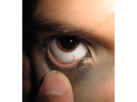
Figure 1
External photograph of right periocular region of Patient 1 demonstrating blue discoloration of skin, conjunctiva, and caruncle.
|
|

Figure 2
T1-weighted magnetic resonance imaging (MRI) of patient 1 demonstrating extensive disease (white arrow) within the right orbit extending to the orbital apex and through the inferior orbital fissure to the pterygopalatine fossa.
|
|
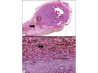
Figure 3
A, Histologic sections from resection specimen from patient 1 reveal a monotonous proliferation of malignant epithelioid cells forming a well-circumscribed nodule in association with a blue nevus (outlined section indicates the area magnified in part). B, Atypical cellular blue nevus component is designated by the solid black arrow, while the invasive melanoma forming an expansile nodule is designated by the solid white arrow. Malignant epithelioid cells (solid white arrow) juxtaposed with benign spindled blue nevus component (solid black arrow).
|
|
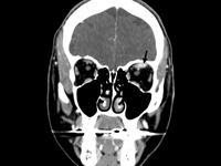
Figure 4
Computed tomography scan of patient 2 demonstrating a mass in the left superior orbit infiltrating the superior rectus muscle (black arrow).
|
|
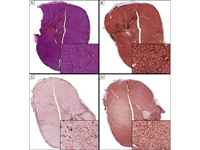
Figure 5
Histologic sections of resection specimen from patient 2 revealing a monotonous proliferation of malignant epithelioid cells forming a well-circumscribed nodule in fibrovascular soft tissue (A). Immunohistochemical studies demonstrate diffuse, strong positivity in the tumor cells for markers of melanocytic differentiation, including Melan-A (B), S100 (C), and HMB-45 (D). The combined positivity for these markers confirms melanocytic differentiation by the tumor cells and, together with the morphology, confirms the diagnosis of melanoma.
|
|

Figure 6
Axial T2-weighted MRI of patient 3 demonstrating a mass medial to the optic nerve and just posterior to the globe on the right side (white arrow).
|
|

Figure 7
A, Histologic sections of resection specimen from patient 3 reveal a proliferation of spindle and epithelioid cells forming an expansile nodule in fibrovascular soft tissue with tumor necrosis present in the area designated by the asterisk; the dashed line designates the boundary between viable and necrotic tumor. B, Perineural invasion is designated by the solid white arrow. C, High-power examination reveals proliferation of malignant epithelioid melanocytes forming an expansile nodule (solid black arrow), which is juxtaposed with a proliferation of cytologically benign spindled and dendritic melanocytes (designated by a solid white arrow).
|
|
| Discussion | We highlight the importance of careful evaluation of surgical biopsy specimens for detection of associated cellular blue nevus in patients with primary orbital melanoma. The finding of a cellular blue nevus supports the diagnosis of a primary orbital melanoma rather than a metastatic lesion. The identification of a cellular blue nevus component in the orbital specimen together with a negative systemic work-up, which should include at least a total body PET-CT scan and a total body skin check, gives high likelihood for the diagnosis of a primary rather than a metastatic orbital melanoma.
The histologic diagnosis of a blue nevus or a cellular blue nevus can be difficult because of the relative rarity of these lesions. In addition, whereas the invasive melanoma component may involve a large component of the orbital mass, the blue nevus component may represent a small part of the surgical biopsy specimen and could be overlooked. The correct diagnosis may also depend on the experience of the pathologist.
The case of patient 1 highlights the possibility of malignant transformation and the importance of surveillance in patients with congenital blue nevus involving the periocular skin. This type of transformation has previously been described in the ophthalmic literature.(4) In a patient with congenital blue nevus, any changes in size or appearance of the nevus and any onset of proptosis or displacement of the globe should prompt a biopsy of the involved area to rule out transformation to invasive melanoma.
Orbital melanoma usually metastasizes hematogenously, unlike eyelid or conjunctival melanoma, which metastasizes via the draining lymph nodes. This metastatic behavior of orbital melanoma is presumed to be due to the lack of lymphatics in the orbit. After treatment of a primary orbital melanoma in a patient with blue nevus follow-up surveillance might include total body PET-CT scans and careful clinical examinations. | | | Acknowledgements | Supported in part by the National Cancer Institute under award number P30CA016672.
Presented at the Fall 2011 meeting of the ASOPRS, Orlando, Florida. | | | References | 1. Shields CL, Shields JA. Ocular melanoma: relatively rare but requiring respect. Clin Dermatol 2009;27:122-33.
2. Tellado M, Specht CS, McLean IW, Grossniklaus HE, Zimmerman LE. Primary orbital melanomas. Ophthalmology 1996;103:929-32.
3. Polito E, Leccisotti A. Primary and secondary orbital melanomas: a clinical and prognostic study. Ophthal Plast Reconstr Surg 1995;11:169-81.
4. Gündüz K, Shields JA, Shields CL, Eagle RC Jr. Periorbital cellular blue nevus leading to orbitopalpebral and intracranial melanoma. Ophthalmology 1998;105:2046-50.
5. Rice CD, Brown HH. Primary orbital melanoma associated with orbital melanocytosis. Arch Ophthalmol 1990;108:1130-4.
| |
|
|
|
|
|
|
 Welcome, please sign in

