|
|
 |
|
|
|
|
|
Keratouveitis Following A Contralateral Herpes Zoster Ophthalmicus
Digital Journal of Ophthalmology 2007
Volume 13, Number 1
July 9, 2007
|
Printer Friendly
|
|
|
|
|
| Abstract | Objective
To report a patient who had previous unilateral herpes zoster ophthalmicus presenting with contralateral ocular involvement.
Methods
Case report
Results
A 47 year-old woman presented with a unilateral, painful, red eye preceded by a painful rash over her contralateral forehead and nose. Examination revealed a healed herpes zoster rash on the right forehead and tip of the nose associated with left eye keratouveitis. Examination of the right eye did not reveal any abnormality despite being on the same side with the facial scar. She was treated with topical antiviral drops with subsequent topical steroid. After one week of treatment, vision improved to 6/12. She had completely recovered one month later. A faint corneal scar was present inferonasally that did not involve visual axis. The final visual acuity in the left eye was 6/9.
Conclusion
We propose a careful ophthalmic examination in both eyes in patients with herpes zoster ophthalmicus in view of the possibility of contralateral eye involvement.
Keywords
Contralateral, Herpes Zoster Ophthalmicus, Keratouveitis | | | Introduction | | Involvement of the ophthalmic branch of trigeminal nerve by herpes zoster can cause various ocular complications. We report a patient who had unilateral herpes zoster ophthalmicus presenting to us with contralateral ocular involvement. | | | Materials and Methods | | Case report | | | Results | A 47 year-old woman was referred for unilateral painful red eye associated with a decrease in vision. The symptoms were preceded by a painful rash over her contralateral forehead and nose one week earlier. She was an otherwise healthy lady with no known medical problems.
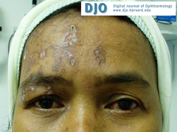
At presentation, there was a facial scar on the right forehead and the tip of nose corresponding to the dermatomal distribution of the ophthalmic division of the trigeminal nerve. The lids were spared. (Figure 1) Examination of the left eye revealed vision of 6/60, with pinhole improvement to 6/36. The corneal sensation was decreased. There were paracentral Descemet’s folds at the 5 o’clock position, measuring 3mm by 3mm with very mild stromal edema, endothelial bullae and localized keratic precipitates. The anterior chamber showed mild inflammation. The pupil was otherwise reactive and round. (Figure 2) The intraocular pressures and ocular movement in both eyes were normal. Visual acuity in the right eye was 6/9. The examination of the right eye did not reveal any abnormality despite being on the same side with the facial scar.
The distribution of the facial scar was characteristic of herpes zoster ophthalmicus (HZO) with a possible relation to the left eye keratouveitis. Topical antiviral (Acyclovir) with subsequent topical steroid were prescribed. She was also given analgesia for residual right sided facial pain. After one week of treatment, the vision improved to 6/12 in the left eye. The corneal edema and striae were markedly reduced and the bullae the on endothelium had completely resolved.
She completely recovered one month later. There was no more facial pain. A faint corneal scar was present inferonasally that did not involve the visual axis. The final vision in the eye left was 6/9.
| | | Discussion | Herpes zoster, commonly known as shingles, is caused by reactivation of latent varicella-zoster virus (VZV) present in sensory ganglia after chicken pox infection.(1) Any sensory ganglion may be involved but the dermatomes most commonly affected, in order of frequency, are thoracic, cranial, cervical, lumbar and sacral.(1, 2) Herpes Zoster Ophthalmicus (HZO) involves the dermatome innervated by the ophthalmic division of the trigeminal nerve and accounts for 20% of all cases of Herpes Zoster.(3)
HZO differs from other types of herpes zoster because of the likely involvement of the globe itself as opposed to herpes zoster in other dermatomes which is typically limited to localized cutaneous disease. Corneal involvement may complicate HZO in 34% of cases. It can manifest concurrently with the disease or present later. One such example is keratouveitis which has a typical onset of seven days after HZO.(1, 2, 4)
Nakanishi et al. reported a patient who developed acute retinal necrosis (ARN) in the contralateral eye following HZO.(5) There was also a reported case of an immunocompromised patient who had a unilateral HZO with ipsilateral ocular blindness and subsequently developed encephalitis and antegrade spread of VZV through the visual system resulting in bilateral blindness.(6) However, to the best of our knowledge, keratouveitis in contralateral eye following HZO has not been previously reported.
The possible explanation for corneal involvement in our patient is direct cytopathic effect of the virus and corneal immune response to replicating viral particles.(1, 2) The pathogenesis, however, for contralateral ocular involvement is obscured. Our patient might have had a bilateral HZO with insufficient dermal findings on the left side.
In most situations, diagnosis of HZO can be established from the characteristic rash.(1) Invasive laboratory investigations are reserved only for those atypical cases of HZO.(1) In our patient, they were obviously contraindicated in view of the presence of a good visual potential and good response to treatment.
Apart from presenting early during the course of the disease, grave complications such as ARN, meningitis, encephalitis and cerebral infarct may occur later hence the need for an appropriate follow up for patients with HZO.(1, 5, 6)
| | | References | 1. Krachmer, Mannis, Holland. Cornea and External Disease: Clinical Diagnosis and Management. Cornea: Mosby, 1997:1225-1242.
2. Looney BD. Herpes Zoster Ophthalmicus. Clinical Eye and Vision Care 1997;9:203-211.
3. Rogazzino MW, Melton, Kurland LT. Population-based study on herpes zoster and its sequela. Medicine 1982;61:310.
4. Marsh K, Cooper M. Ophthalmic herpes zoster. Eye 1993;7:350-370.
5. Nakanishi F, Takahash H, Ohara K. Acute Retinal Necrosis Following Contralateral Herpes Zoster Ophthalmicus. Japanese Journal of Ophthalmology 2000;44(5):561-564.
6. Rostad SW, Olson K, McDougall J, Shaw C-M, Alvord EC. Transsynaptic spread of varicella zoster virus through the visual system: A mechanism of viral dissemination in the central nervous system. . Human Pathology 1989;20(2):174-179.
| |

Figure 1
Facial scar at the dermatomal distribution of ophthalmic division of right trigeminal nerve
|
|

Figure 2
Left eye keratouveitis
|
|
|
|
|
|
|
|
 Welcome, please sign in