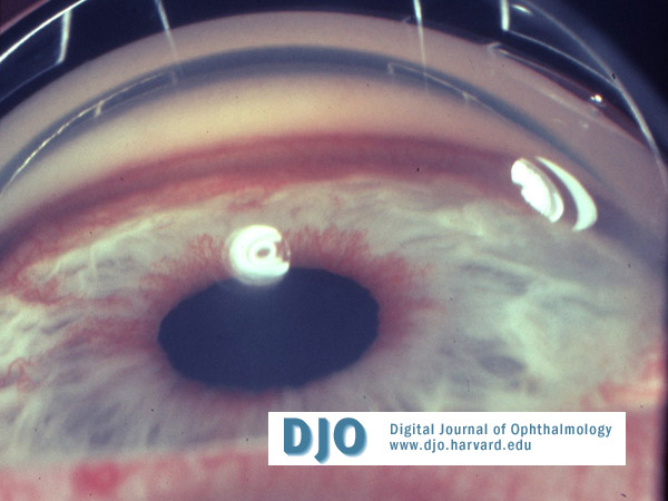
Case: A 72-year-old female with diabetes complained of decrease in vision and redness of her right eye. Best corrected visual acuity OD was count fingers at 2 feet and OS 20/80. The intraocular pressure OD was 40 mmHg (Figure). The anterior segment exam of the other eye was notable only for a moderate nuclear sclerotic cataract. Gonioscopy OD was notable for an open angle with 360º of neovascularization. Gonioscopy OS revealed an angle open to ciliary body 360º. Dilated fundus exam revealed proliferative diabetic retinopathy OU.
 Welcome, please sign in