Carmen Gonzales, MD | Massachusetts Eye and Ear Infirmary, Harvard Medical School Lois Hart, RDMS | Massachusetts Eye and Ear Infirmary, Harvard Medical School
Figure 1
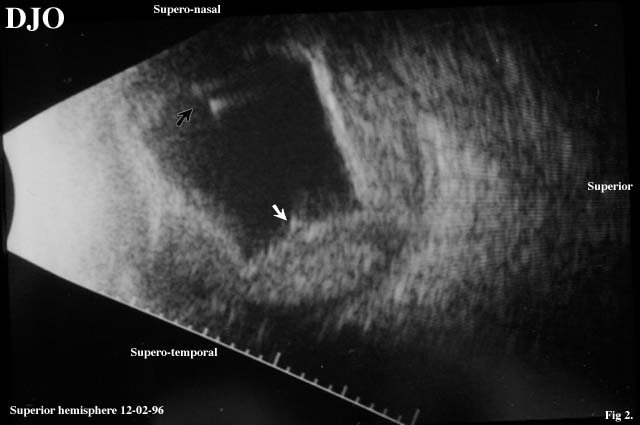
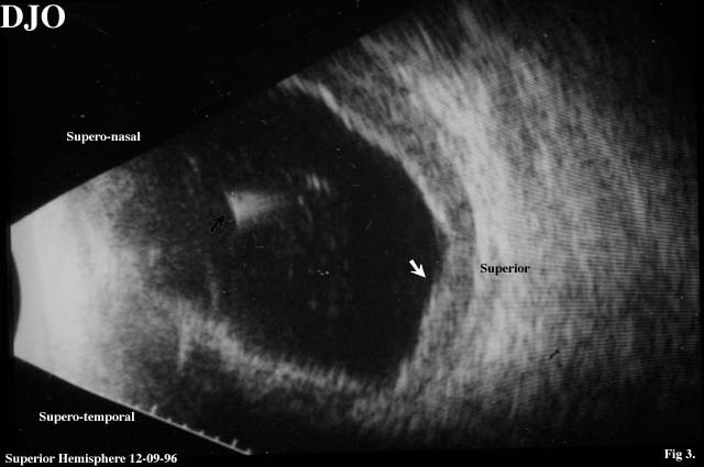
Figures 1-3. This is ultrasound of a patient with a history of a ruptured globe OD. Ultrasound is requested to rule out posterior segement pathology. B-scan ultrasound was performed 2 and 9 days post ruptured globe repair.
Figure 2
Figure 3
Questions
1. Describe the findings of the B-scan ultrasound
2. What is the significance of the intravitreal echoes (black arrows figures 2 and 3)?
3. Could these intravitreal echoes represent air bubbles?
4. What is the significance of the low to moderate amplitude echoes posterior to the scalloped shaped elevations?
5. Based on the ultrasound appearance, how would you manage this patient?
 Welcome, please sign in