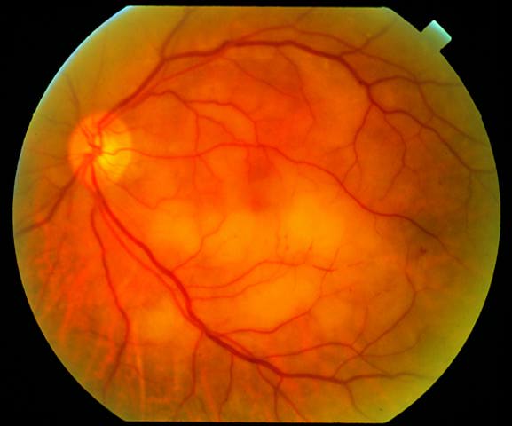
Figure 1
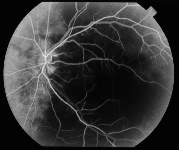
Figures 1-3. This patient is an elderly man who had sudden visual loss in his left eye. On testing, the patient was no light perception OS. Figure 1 shows the patient's fundus OS. There is retinal edema in the macula and subretinal pallor underneath the inferior arcade. A cherry red spot is seen at the fovea. Figures 2 and 3 SHOW fluorescein angiograms of OS. There was no arterial filling noted until 31 seconds. At 40 seconds the arteriovenous phase begins. Patchy hypofluorescence of the retina and choroid is seen. At 720 seconds perivascular staining is observed. There is also subretinal hyperflourescence with leakage.
Figure 2
Figure 3
Questions
1. What is your differential diagnosis?
2. What is the most likely diagnosis?
3. How common is a cherry red spot in this condition?
4. How can electroretinography be helpful in the diagnosis of this condition?
5. What is the treatment for this conditon and what is its prognosis?
 Welcome, please sign in