|
|
 |
|
|
|
|
|
Neuro-ophthalmology Quiz 6
|
Printer Friendly
|

Howard Pomeranz, M.D., Ph.D | Massachusetts Eye and Ear Infirmary, Harvard Medical School, University of Maryland Medical Center M.A. Afshari, M.D., M.P.H. | Massachusetts Eye and Ear Infirmary, Harvard Medical School August 2, 1997
|
|
[View Answers] [Back to Neuro-ophthalmology]
|
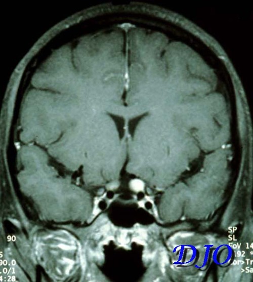
Figure 1
Brain MRI (Coronal). MRI scan of the brain revealed enlargement of the left optic nerve and enhancement of the left optic nerve with gadolineum, as illustrated in the coronal section below.
|
| | Case History | A 23 year-old woman complains of "fading in and out " of her vision in the left eye several times a day for two weeks accompanied by mild headache. A CT scan of the head was negative. She was treated with analgesics and acupuncture with improvement in the headaches. The patient subsequently developed pain with eye movement, intermittent numbness and tingling of the left leg and noted gradual worsening of her visual acuity until able to see only "a sliver of vision" in the left eye. The following day she developed complete loss of vision in the left eye. Family history and the patient's medical history were noncontributory.
On examination visual acuity was 16/20 OD and no light perception OS. There was an afferent pupillary defect on the left. The left pupil was sluggishly responsive to consensual light reflex. The exam was otherwise remarkable for a swollen optic nerve on the left. | | | Questions | 1. What is the diagnosis and what test might be suggested?
2. What treatment options could be presented to the patient?
| | | [View Answers] |
|
|
|
|
|
|
 Welcome, please sign in


