Howard Pomeranz, M.D., Ph.D. | Massachusetts Eye and Ear Infirmary, Harvard Medical School, University of Maryland Medical Center M.A. Afshari, M.D., M.P.H. | Massachusetts Eye and Ear Infirmary, Harvard Medical School
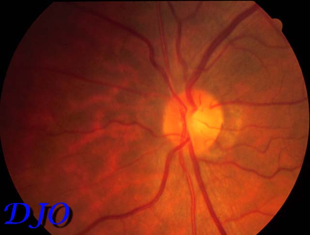
A 61 year-old Korean man was in good health until 2 years prior to presentation when he noted blurriness in the right eye, making it difficult for him to read. This was not helped by a change in eyeglass prescription. The vision continued to deteriorate in the right eye until one year ago when vision was totally lost. Review of past medical and ocular histories was noncontributory. On examination at this time his visual acuities were no light perception OD and 16/13 OS. The right pupil was mid-dilated and an afferent pupillary defect was present. There was no proptosis, ptosis or motility deficit present. There was no dyschromatopsia or metamorphopsia present in the left eye. Neurological exam was unremarkable. Visual field was full on the left eye. Fundus findings are illustrated below.
 Welcome, please sign in