|
|
 |
|
|
|
|
|
Retina/Uveitis Quiz 24: A 60-year-old man with a progressive, bilateral, vision loss
|
Printer Friendly
|
 Lily S. Ooi, MBBS
 | Princess Alexandra Hospital Mark Donaldson, FRANZCO | Princess Alexandra Hospital, Brisbane, Queensland, Australia February 3, 2011
|
|
[View Answers] [Back to Retina/Uveitis]
|

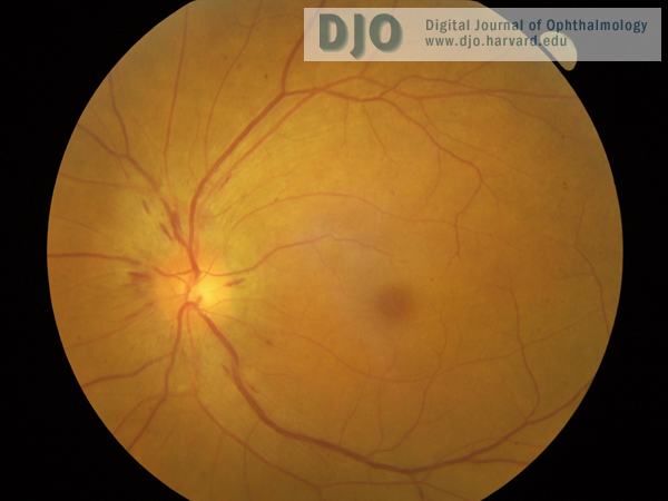
Figure 1
Left fundus showing disc swelling with nerve fiber layer hemorrhages.
|

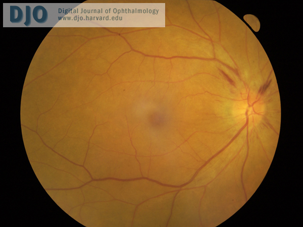
Figure 2
Right fundus, with similar appearance to left fundus.
|
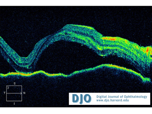
Figure 3
OCT of right macula (left macula has similar appearance).
|
| | Case History | | A 60-year-old Vietnamese man presented with a 2-week history of progressive bilateral vision loss associated with bilateral hearing loss. He had a history of well controlled diabetes and ischemic heart disease. There was no other relevant ocular history, and he was otherwise in good health. Best-corrected visual acuity was 20/60 in both eyes. Slit-lamp examination showed bilateral anterior uveitis, vitritis, disc swelling with nerve fiber layer hemorrhages (Figures 1 and 2), and multifocal subretinal exudative detachments (Figure 3). | | | Questions | 1. What is the differential diagnosis?
2. What further history or investigations are necessary to make the diagnosis?
3. What is the most likely diagnosis?
4. What are the clinical features of this condition?
5. What is the treatment? | | | [View Answers] |
|
|
|
|
|
|
 Welcome, please sign in