|
|
 |
|
|
|
|
|
Cornea/Refractive Surgery Quiz 25: A 61-year-old man with a complication of intraocular surgery
|
Printer Friendly
|
 Tarek Alasil
 | Doheny Eye Institute, University of Southern California Michael Engelbert | Edward S. Harkness Eye Institute, lumbia University Lama A. Al-Aswad | Edward S. Harkness Eye Institute, Columbia University September 5, 2008
|
|
[View Answers] [Back to Cornea/Refractive Surgery]
|

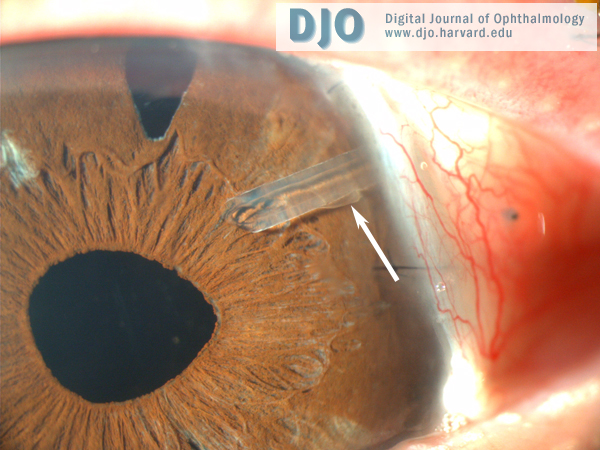
Figure 1
Slit-lamp examination of the left eye shows an iatrogenic Descemet’s membrane tear (arrow). There is no corneal edema noted.
|

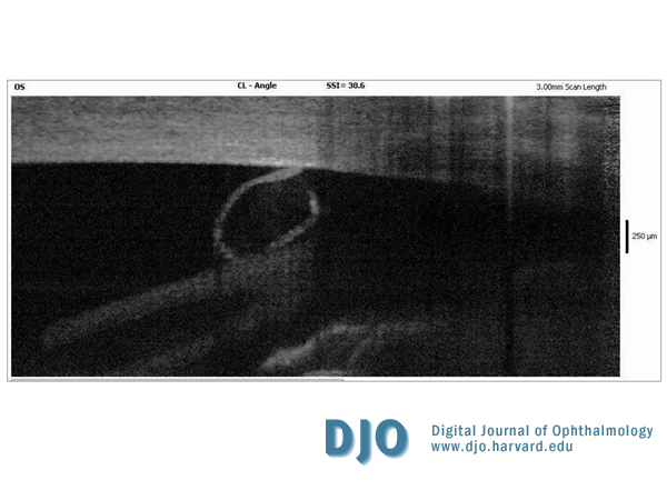
Figure 2
Anterior segment optical coherence tomography of the left eye showed the Ahmed valve tube and iatrogenic Descemet’s membrane tear. There is no corneal edema noted.
|
| | Case History | | A 61-year-old man with advanced open angle glaucoma and visually significant cataracts underwent combined cataract extraction, intraocular lens implantation, and Ahmed valve placement in the left eye. Six weeks later, the patient was evaluated in the clinic for a routine follow-up. The slit-lamp exam and anterior segment optical coherence tomography done at that time are shown below. | | | Questions | 1. What condition does this patient have?
2. How did the patient develop this condition, and what is its clinical significance?
3. At which point during glaucoma or cataract surgery can the Descemet's membrane be torn or stripped?
4. How could you manage the Descemet’s tear intraoperatively in order to avoid further stripping of the membrane?
5. What should you do when closing the incision, and why?
6. What other modalities can you consider for fairly large Descemet’s detachments?
7. How does gas treat a Descemet’s detachment? | | | [View Answers] |
|
|
|
|
|
|
 Welcome, please sign in