Coats disease mimicking retinoblastoma
Feb 20, 2022
Volume 28, Number 1
Dolores Ríos y Valles-Valles, MD | Ophthalmic Pathology Department, Asociación para Evitar la Ceguera en México I.A.P.
Ivette Hernández-Ayuso, MD | Ophthalmic Pathology Department, Asociación para Evitar la Ceguera en México I.A.P.
Alejandra Lozano Bustillo, MD | Ophthalmic Pathology Department, Asociación para Evitar la Ceguera en México I.A.P.
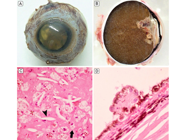
A 3-year-old girl presented at Asociación para Evitar la Ceguera en México with leukocoria and a blind and painful left eye. Ultrasound revealed a fully occupied vitreous cavity with calcifications and total retinal detachment. Enucleation was performed due to suspected retinoblastoma. Gross findings showed an extensive, yellow, subretinal exudation throughout the vitreous chamber, with the retina displaced anteriorly (A-B). Histopathologically, abundant slit-like cholesterol clefts (arrow head) and foamy histiocytes (arrow) were seen (C), consistent with Coats disease. In addition to these findings, distrophic calcium salts deposits and congenital drusen were identified (D). Coats disease is diagnosed as a different entity in nearly 50% of cases. It is frequently mistaken for retinoblastoma.
 Welcome, please sign in


