Persistent fetal vasculature on RetCam
Dec 31, 2020
Volume 26, Number 4
Ghazal Valizadeh, BMedSt | Bond University, Gold Coast, Queensland, Australia
Ashely J. Porter, BMed, MMed | Department of Ophthalmology, Gold Coast University Hospital, Gold Coast, Queensland Australia
Heather C. Russell, MbChb (Hons), BSc (Hons), FRCOphth, FRANZCO | Department of Ophthalmology, Gold Coast University Hospital, Gold Coast, Queensland, Australia
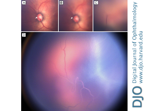
A premature neonate, born at 27 weeks’ gestational age and weighing 900 g, was referred for retinopathy of prematurity screening at Gold Coast University Hospital. The child was delivered by emergency cesarean section for fetal distress in the setting of maternal pyrexia. On RetCam imaging (Natus Medical Inc, Pleasanton, CA) at 36 weeks, a persistent hyaloid artery was seen extending from the optic nerve to the posterior lens surface, indicating persistent fetal vasculature (A-C). The branching vessels from the hyaloid artery were seen extending over the posterior surface of the lens capsule, with evidence of intravascular blood flow (D). There was no evidence of cataract, microphthalmia, or retinopathy of prematurity. Ongoing follow-up was arranged to monitor for cataract development.
 Welcome, please sign in


