Ciliary body metastasis from prostate adenocarcinoma completely resolves with systemic therapy
Dec 23, 2020
Volume 26, Number 4
Catherine M. Marando, MD | Department of Ophthalmology, Massachusetts Eye and Ear, Boston
Nazlee Zebardast, MD, MSc | Department of Ophthalmology, Massachusetts Eye and Ear, Boston
Mary E. Aronow, MD | Department of Ophthalmology, Massachusetts Eye and Ear, Boston
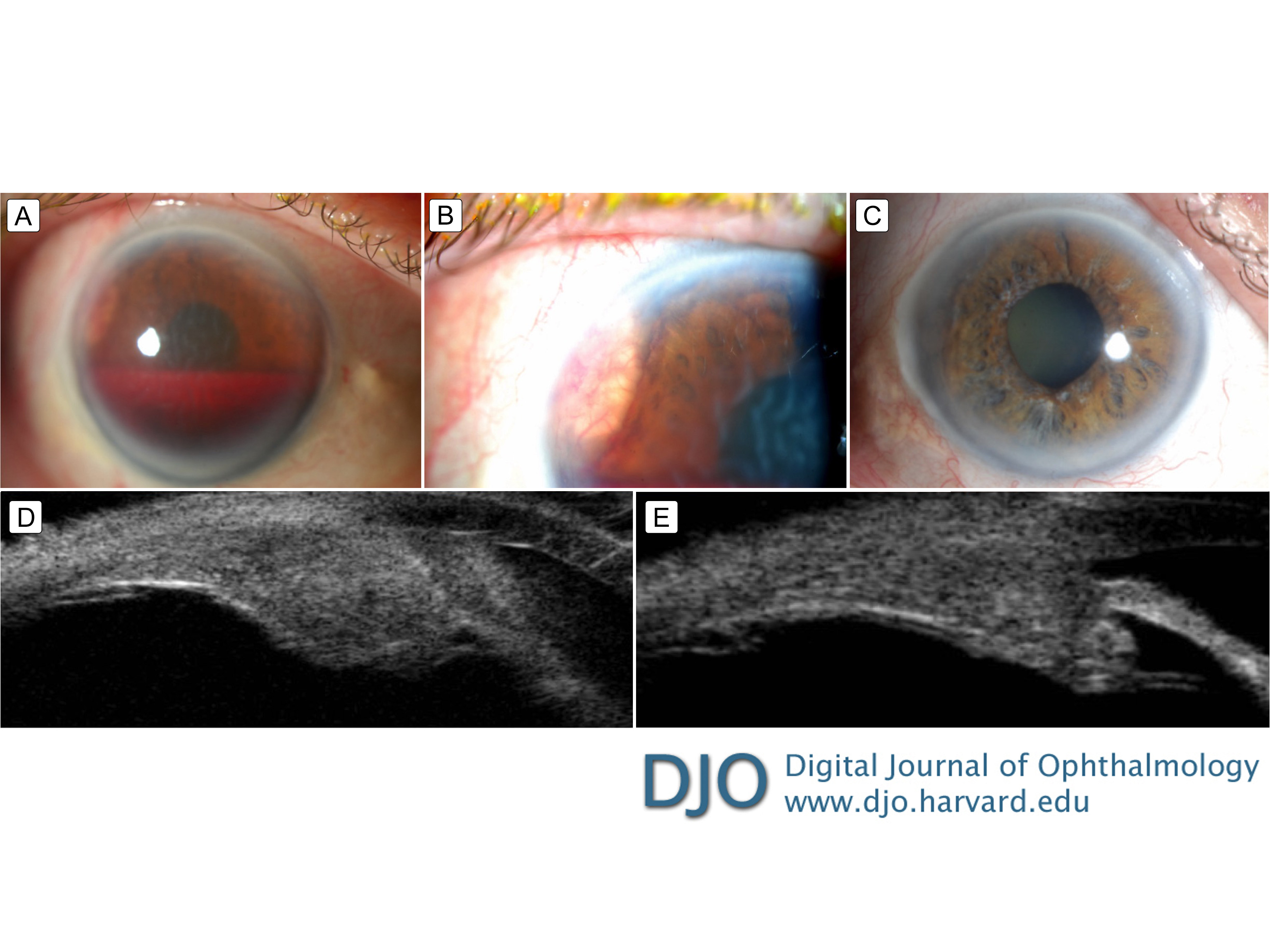
A 68-year-old man presented emergently at Massachusetts Eye and Ear with acute pain and decreased vision in his right eye. On examination, visual acuity in the right eye was light perception; intraocular pressure (IOP) measured 61 mm Hg. Slit-lamp examination demonstrated total hyphema and a whitish-tan vascular lesion in the anterior chamber angle at 10 o’clock. Three days later, when the hyphema began to clear, the vascular lesion became more apparent (A-B). Ultrasound biomicroscopy (UBM) revealed a ciliary body mass with extension into the angle. No choroidal lesions were identified on fundus examination or ultrasonography. Topical ocular antihypertensive therapy and intravenous acetazolamide were administered with an acceptable decrease in IOP. Computed tomography of the chest, abdomen, and pelvis showed diffuse lytic and sclerotic bony metastases, and biopsy of a bony mass confirmed metastatic prostate adenocarcinoma. The patient was started on androgen deprivation therapy and docetaxel. Six months later, his visual acuity had returned to 20/20, and the ciliary body metastasis demonstrated complete regression on slit-lamp examination (C), which was confirmed by comparing the UBM on initial presentation (D) to the UBM 6 months after treatment (E).
 Welcome, please sign in