|
|
|
Register
with DJO to receive personalized updates.
If you're already a
member, please sign in.
|
|
|
|
|
|
|
|
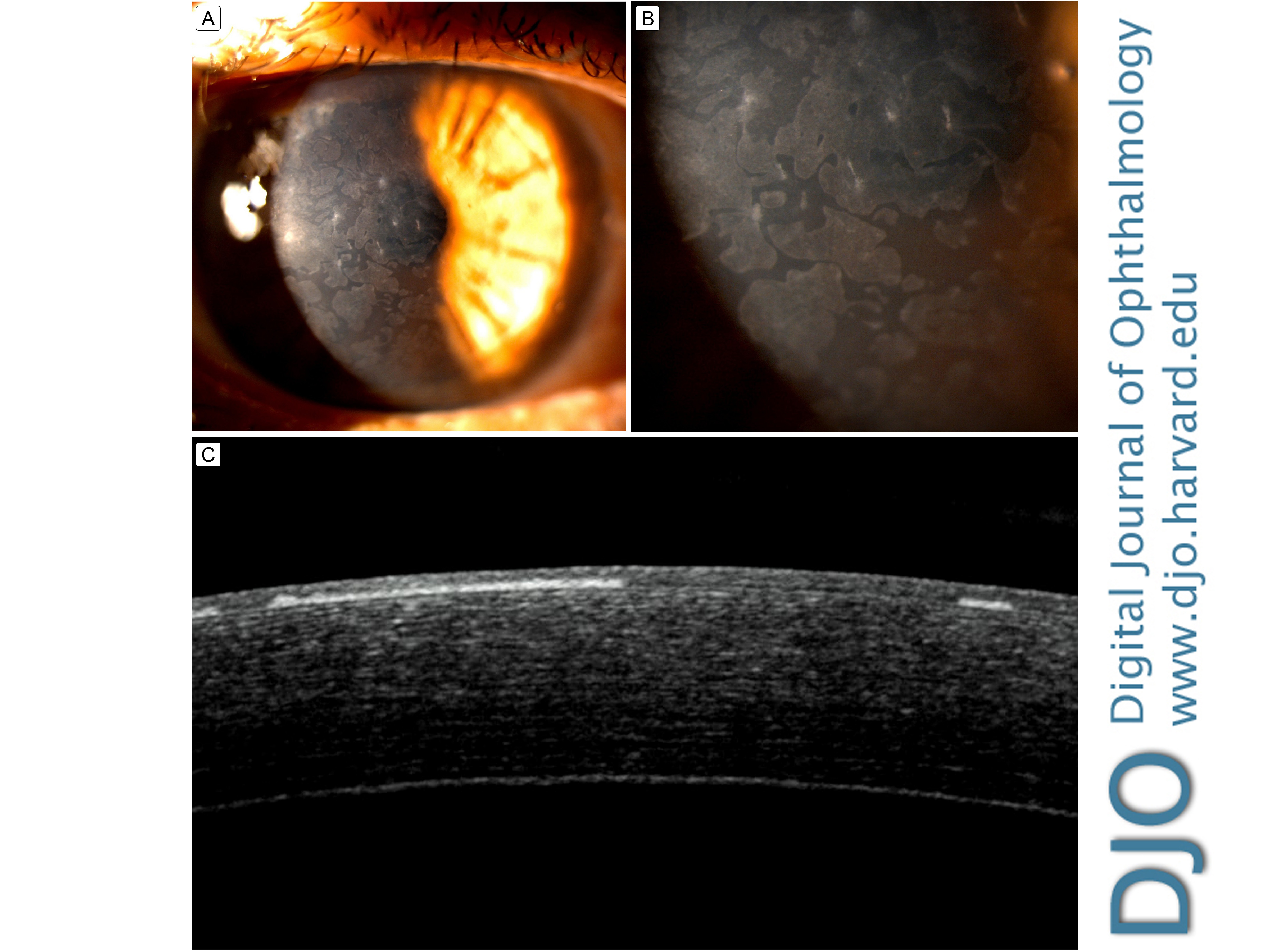
Atypical presentation of unilateral map-dot dystrophy Feb 20, 2022 Volume 28, Number 1 Sudhakar Potti, MS, FLVPEI | Department of Cornea and Refractive Surgery, Sankara Eye Hospital, Guntur, Andhra Pradesh, India Aparna N. Nayak, MS, DNB, FICO | Department of Cornea and Refractive Surgery, Sankara Eye Hospital, Guntur, Andhra Pradesh  A 60-year-old woman presented at Sankara Eye Hospital, Guntur, Andhra Pradesh,India, with foreign body sensation and watering in her right eye. She had undergone cataract surgery 6 months earlier. Medical history included diabetes and hypertension, for which she was taking medications. On examination, best-corrected visual acuity was 6/24 in the right eye and 6/12 in the left eye. Slit-lamp examination showed multiple maplike subepithelial lesions with well demarcated margins spread diffusely on the cornea, with dispersed filaments (A, B). Her eye had a clear cornea, with senile cataract. Anterior segment optical coherence tomography (AS-OCT) showed well defined hyper-reflective lesions limited to the basement membrane (C). Dry eye evaluation was normal. A diagnosis of unilateral map-dot dystrophy was made based on clinical morphology and confirmed by AS-OCT. |
|
|
|
|
|
 Welcome, please sign in