
|
|
|
Register
with DJO to receive personalized updates.
If you're already a
member, please sign in.
|
|
|
|
|
|
|
|
Choriocapillaris nonperfusion in association with choroidal nodules in a patient with neurofibromatosis type 1 depicted on optical coherence tomography angiography Aug 11, 2021 Volume 27, Number 3 Mehreen Adhi, MD | Department of Ophthalmology and Visual Sciences, Louisiana State University, New Orleans Maria Reinoso, MD | Department of Ophthalmology and Visual Sciences, Louisiana State University, New Orleans 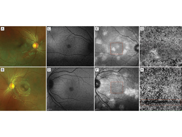 A 14-year-old girl presented at Louisiana State University Health Care Network for retina evaluation. She had a known diagnosis of neurofibromatosis type 1. She had inoperable plexiform nerofibromas of the brain and was being considered for treatment with selumetinib, a mitogen-activated protein kinase enzyme inhibitor. Her ocular history was significant for barrier laser retinopexy for a peripheral retinal hole in the right eye and a retinal tear in the left eye. On examination, her best-corrected visual acuity was 20/20 in the right eye and 20/25 in the left eye, with no improvement on refractive correction. Anterior segment examination of both eyes was normal. Dilated fundus examination of the posterior pole and midperiphery was normal in both eyes. In the far periphery, there were laser scars barricading a retinal hole in the superior periphery in the right eye and a retinal tear in the inferior periphery in the left eye. Color fundus photographs and fundus autofluorescence were normal in both eyes (A-D). Near infrared scanning laser ophthalmoscope imaging demonstrated large, symmetric, placoid-type lesions consistent with choroidal nodules in both eyes (E-F). Of note, en face spectral domain optical coherence tomography (SD-OCT) angiography (Heidelberg Spectralis, Heidelberg, Germany) at the level of choriocapillaris demonstrated areas of nonperfusion in both eyes (G-H). These areas of nonperfusion appeared to be exaggerated adjacent to the placoid-type choroidal nodules when correlated with near infrared scanning laser ophthalmoscope imaging (E and F, resp.). This en face SD-OCT angiography finding could represent either thinning or disruption and/or focal atrophy of the choriocapillaris induced by presence of the adjacent long-standing placoid-type choroidal nodules. Note, the boxed area of H depicts a region of motion artifact in the en face SD-OCT angiography image; hence, choriocapillaris nonperfusion in this region should be interpreted with caution. |
|
|
|
|
|
 Welcome, please sign in


