
|
|
|
Register
with DJO to receive personalized updates.
If you're already a
member, please sign in.
|
|
|
|
|
|
|
|
Melanin-producing central ulcer caused by an Exophiala species Jan 13, 2020 Volume 26, Number 1 Shyam A. Patel, MD, MBA | Eye Consultants of Atlanta, Atlanta, Georgia Charles D. Salisbury, MD | Eye Consultants of Atlanta, Atlanta, Georgia Stephen M. Hamilton, MD | Eye Consultants of Atlanta, Atlanta, Georgia 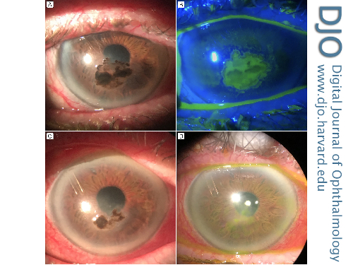 A 91-year-old woman, with an ocular history of a neurotrophic cornea and neovascular glaucoma secondary to a retinal vein occlusion, presented at Eye Consultants of Atlanta with a corneal ulcer in her right eye. The ulcer consisted of dense, brown pigmentation and an epithelial defect centrally, confirmed by fluorescein staining (A, B). Fungal culture grew an Exophiala species, which are black yeast that can cause, in addition to keratitis, multiple types of infections, including central nervous system and respiratory infections. The cell walls of these organisms contain abundant melanin, which is thought to be protective against both host defenses and antifungal therapy. Natamycin eye drops were started hourly. There was reduction in melanin on examination 9 days after initiation of treatment (C) and no melanin pigment and only trace corneal scarring on follow-up at 1 month (D). Because of glaucoma and vein occlusion, visual acuity at all follow-up examinations was hand motions. |
|
|
|
|
|
 Welcome, please sign in


