Preserved unilateral macular tissue within large geographic atrophy sparing vision
Dec 23, 2019
Volume 25, Number 4
Raviv Katz, MS, BS | Harvard Retinal Imaging Lab, Massachusetts Eye and Ear, Boston, Massachusetts Department of Ophthalmology, Harvard Medical School, Boston, Massachusetts
Grace Kromm | Department of Ophthalmology, Harvard Medical School, Boston, Massachusetts
Archana Nigalye, MD | Department of Ophthalmology, Harvard Medical School, Boston, Massachusetts
John B. Miller, MD | Retina Service and Harvard Retinal Imaging Lab, Massachusetts Eye and Ear, Boston, Massachusetts Department of Ophthalmology, Harvard Medical School, Boston, Massachusetts
Deeba Husain, MD, PhD | Retina Service and Harvard Retinal Imaging Lab, Massachusetts Eye and Ear, Boston, Massachusetts Department of Ophthalmology, Harvard Medical School, Boston, Massachusetts
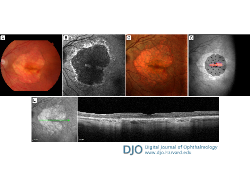
A 92-year-old, otherwise healthy, white woman presented at Massachusetts Eye and Ear with a chief complaint of worsening near vision. She was diagnosed with advanced, atrophic, nonexudative, age-related macular degeneration bilaterally, with subfoveal involvement in the right eye and a thin strip of preserved macular tissue in the left eye. At her recent visit, the patient was newly diagnosed with exudative age-related macular degeneration with active choroidal neovascularization in the left eye. Dilated fundus photographs revealed geographic atrophy (GA) of similar areas in both eyes: 21.777 mm2 in the right eye and 24.197 mm2 in the left eye. Visual acuity was counting fingers at 1' in the right eye and 20/50−2 in the left eye. Fundus examination of the left eye revealed a new hemorrhage at the inferotemporal edge of the GA (A). The band of healthy retinal tissue was oriented in such a way that the geographic atrophy appeared to be in an intriguing pattern of the shape of a heart. This shape is also clearly outlined on autofluorescence and multicolor (B,C). Microperimetry illustrates the remaining functional tissue centrally (D). A representative slice of the Heidelberg OCT illustrates the GA and preserved foveal tissue (E). The reason for this temporal preservation of retinal tissue and development of choroidal neovascularization at the edge of atrophy is not fully understood. Anti-VEGF injection was recommended to preserve vision in her left eye, and the patient received an intravitreal injection of 0.05 cc (1.25 mg) bevacizumab and a second injection 1 month later, with her visual acuity decreasing from 20/50−2 to 20/64+2.
 Welcome, please sign in


