|
|
|
Register
with DJO to receive personalized updates.
If you're already a
member, please sign in.
|
|
|
|
|
|
|
|
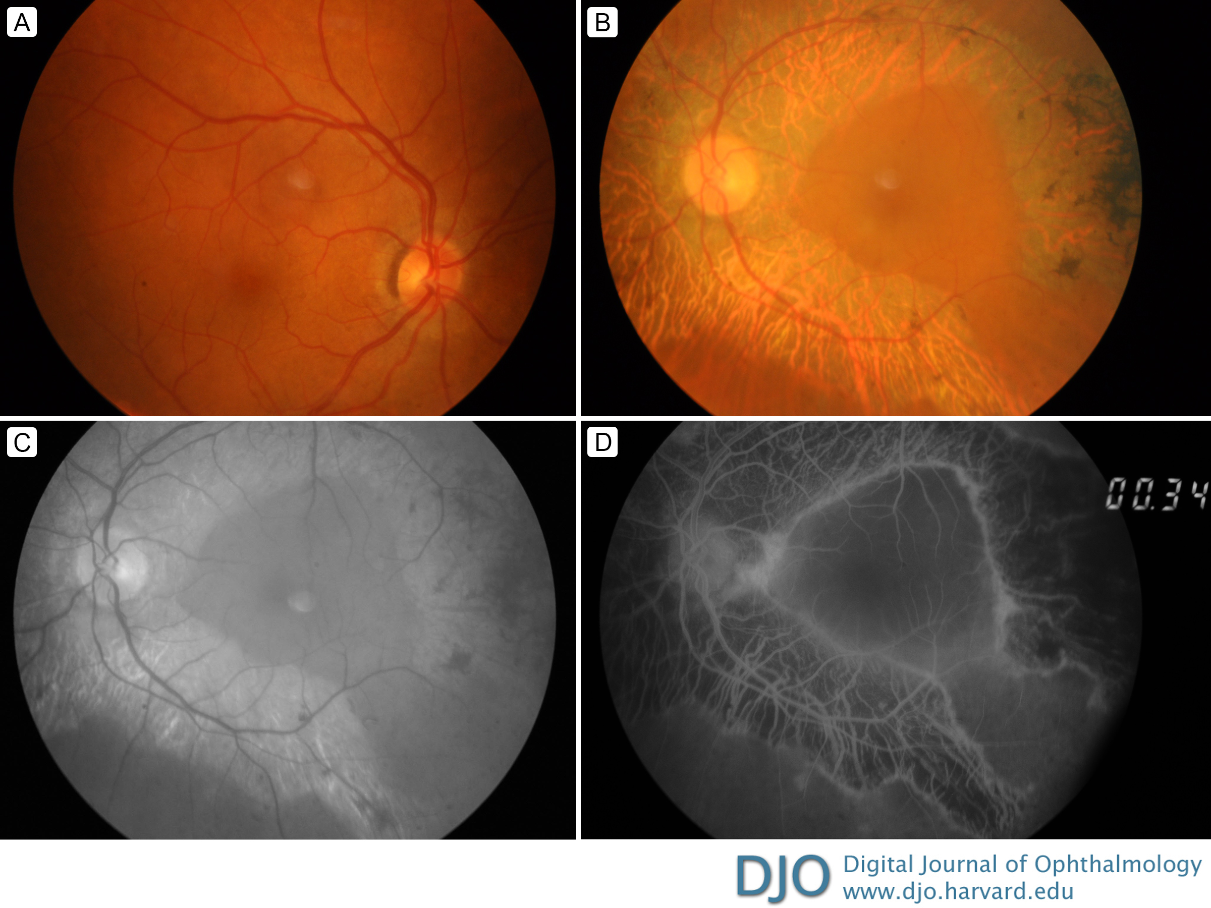
Unilateral pigmented paravenous retinochoroidal atrophy Dec 2, 2019 Volume 25, Number 4 Jorge Moreira, MD | Department of Ophthalmology, Hospital Pedro Hispano, Porto, Portugal Rui Carvaho, MD | Department of Ophthalmology, Hospital Pedro Hispano, Porto, Portugal 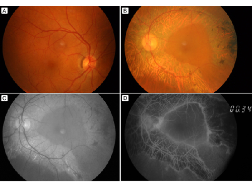 A 55-year-old man presented at the Hospital Pedro Hispano, Porto, with a complaint of low visual acuity in his left eye of several years’ duration. He denied other visual symptoms. His ocular history was unremarkable, and there was no family history of eye disease. On examination, his best-corrected visual acuity was 20/20 in the right eye and 20/50 in the left eye at presentation. Slit-lamp examination revealed a mild cataract in the left eye. Fundus examination of the left eye showed bone-spicule pigmentation associated with retinochoroidal atrophy along the retinal veins, sparing the macula and with no signs of inflammation. Right eye examination was unremarkable (A). The characteristic fundus appearance of the left eye (B) was consistent with a diagnosis of unilateral pigmented paravenous retinochoroidal atrophy. Red-free fundus photography (C) and fluorescein angiography (D, early phase) of left eye highlighted these findings. (In panels A, B, and C there is an artifact in the perifoveal region.) Pigmented paravenous retinochoroidal atrophy is a rare disease of unknown etiology. Patients are usually asymptomatic. The disease is nonprogressive or slowly progressive and is commonly bilateral and symmetric. |
|
|
|
|
|
 Welcome, please sign in