
|
|
|
Register
with DJO to receive personalized updates.
If you're already a
member, please sign in.
|
|
|
|
|
|
|
|
Bergmeister papilla: hollow but not empty May 28, 2019 Volume 25, Number 2 Tarannum Mansoori, MS | Anand Eye Institute, Hyderabad, India 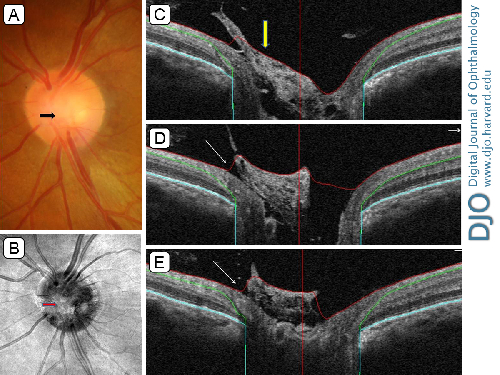 During a routine fundus examination, a 51-year-old man was noted to have grayish-white fibrous tissue on the central surface of the optic nerve head (black arrow), obscuring the cup border, in the left eye (A). A diagnosis of Bergmeister papilla was made. Optical coherence tomography (RTVue XR-100; Optovue, Fremont, CA) confirmed the presence of glial tissue (red arrow) over the optic nerve head in the scanning laser ophthalmoscope image (B). Cross-sectional line scans taken at various depths of optic nerve head (white arrows) revealed tissue in contact with the retinal nerve fiber layer and extending into the vitreous cavity (C-D) and a clear gap between the optic disc and glial tissue and the intact optic disc margin beneath (E, white arrow). |
|
|
|
 Welcome, please sign in


