Wide-field swept source optical coherence tomography angiography: a noninvasive tool for the assessment of retinal changes associated with hypertensive emergency
Jul 22, 2019
Volume 25, Number 3
Yifan Lu, BS | Harvard Retinal Imaging Lab, Massachusetts Eye and Ear, Boston, Massachusetts; Department of Ophthalmology, Harvard Medical School, Boston, Massachusetts
Ying Cui, MD, PhD | Retina Service and Harvard Retinal Imaging Lab, Massachusetts Eye and Ear, Boston, Massachusetts
Ying Zhu, MD | Retina Service and Harvard Retinal Imaging Lab, Massachusetts Eye and Ear, Boston, Massachusetts
John B. Miller, MD | Retina Service and Harvard Retinal Imaging Lab, Massachusetts Eye and Ear, Boston, Massachusetts; Department of Ophthalmology, Harvard Medical School, Boston, Massachusetts
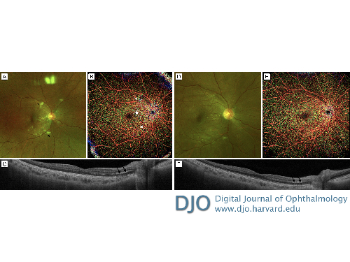
A 29-year-old woman presented at Massachusetts Eye and Ear 2 weeks after experiencing a hypertensive emergency, with blood pressure of 222/144 mm Hg and blurry vision in her left eye. On examination, her uncorrected visual acuity was 20/25 in the right eye and 20/30 in the left eye. Fundus photography (A) showed cotton wool spots (asterisks), macular exudates, and attenuated arterioles. Montage (15 mm × 9 mm) optical coherence tomography (OCT) and OCT angiography (OCT-A; PLEX Elite 9000 Swept-Source OCT; Zeiss, Oberkochen, Germany) demonstrated bilateral optic disc edema, arteriole constriction, and cotton wool spots. The cotton wool spots, located superficially, led to attenuation of the flow signal and downward vertical displacement of the large superficial retinal vessels, which in turn led to segmenting of the typical superficial large retinal vessels more deeply in the retina, producing the intermittent green color (indicating deeper vessels) on depth-encoded OCT-A (B, arrows). Exudates in Henle’s layer of the outer retina are shown on the OCT scan (C, arrows) and the montage OCT-A imaging (B, star). One month after initial presentation, fundus photography and montage OCT-A revealed resolving cotton wool spots (D-E); the exudates in Henle’s layer also showed improvement on OCT imaging (F, arrows). Follow-up OCT-A confirmed that the hypertensive emergency did not lead to permanent retinal vascular damage in this patient. Unlike fluorescein angiography, OCT-A offers a noninvasive tool to identify temporary changes in the retina and to monitor their resolution. This could be particularly important in the assessment of medically unstable patients, as in this case of hypertensive emergency.
 Welcome, please sign in


