Purtscher retinopathy
May 28, 2019
Volume 25, Number 2
Stephen C. Dryden, MD | Hamilton Eye Institute, University of Tennessee Health Science Center, Memphis
Salar Rafieetary, MD | Hamilton Eye Institute, University of Tennessee Health Science Center, Memphis
Lauren C. Ditta, MD | LeBonheur Children’s Hospital, Department of Ophthalmology, Memphis, Tennessee
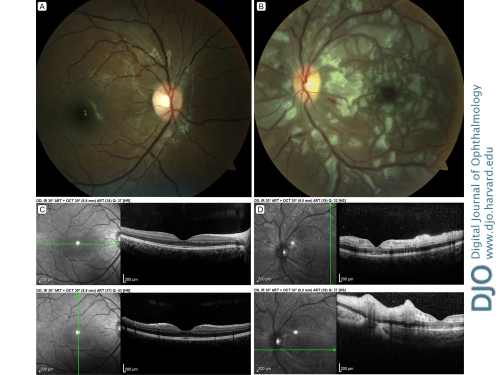
A 12-year-old, African American boy with a past medical history of sickle trait and no past ocular history presented at LeBonheur Children’s Hospital eye clinic in Memphis, Tennessee, with decreased vision in his left eye after a motor vehicle accident. Additional injuries included multiple rib fractures. He denied any direct trauma to his head. His examination was notable for visual acuity of 20/20 in the right eye and counting fingers in the left eye, an afferent pupillary defect in the left eye, and bilateral subconjunctival hemorrhages. Anterior segment examination was otherwise normal. He was found to have a normal-appearing fundus in the right eye (A) and peripapillary and macular cotton wool spots, flame hemorrhages, polygonal areas of retinal whitening and a pseudo-cherry red spot on dilated fundus examination in the left eye (B). Optical coherence tomography revealed a normal macula (C) and hyperreflectivity of the internal and middle layers of the left macula (D). Given the findings on dilated fundus examination and his additional injuries, the patient was diagnosed with Purtscher retinopathy.
 Welcome, please sign in


