Nonsyndromic unilateral posterior lenticonus
Jun 12, 2019
Volume 25, Number 2
Kirandeep Kaur, DNB Ophthalmology | Ophthalmology and Squint Services, Aravind Eye Hospital, Pondicherry, India
Bharat Gurnani, DNB Ophthalmology | Cornea and Refractive Services, Aravind Eye Hospital, Pondicherry, India
Prasanth Gireesh MS Ophthalmology | Cataract and IOL Services, Aravind Eye Hospital, Pondicherry, India
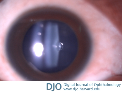
An 18-year-old woman presented at the Aravind Eye Hospital with complaints of painless, gradually progressive diminution of vision in her right eye. On examination, best-corrected visual acuity was 6/9 in the right eye and 6/6 in the left eye. Anterior segment examination was normal in the right eye except for a posterior lenticular bulge, shown in the figure (magnified slit-lamp photograph). Anterior segment examination of the left eye was within normal limits, and posterior segment examination was normal in both eyes. Routine blood investigations were normal. Urine examination was negative for microscopic hematuria and microalbuminuria. Clinical examination excluded sensorineural deafness.
Lenticonus is a cone-shaped deformation of the anterior or, more commonly, the posterior lens capsule. It may be inherited as an autosomal recessive trait or be associated with other abnormalities, such as Alport syndrome or Lowe oculocerebral syndrome (associated with posterior lenticonus). It may occur sporadically. Posterior lenticonus causes lenticular myopia with irregular astigmatism. If the lenticonus is visually significant or if amblyopia is present, lens removal is indicated. In this case, the patient was advised to wear a contact lens in the right eye alone and asked to return for follow-up examination after 6 months.
 Welcome, please sign in


