Avellino (combined granular-lattice corneal) dystrophy
Dec 23, 2018
Volume 24, Number 4
Anchal Thakur, MS | Advanced Eye Centre, Department of Ophthalmology, Postgraduate Institute of Medical Education and Research (PGIMER), Chandigarh, India
Sabia Handa, MS | Advanced Eye Centre, Department of Ophthalmology, Postgraduate Institute of Medical Education and Research (PGIMER), Chandigarh, India
Amit Gupta, MS | Advanced Eye Centre, Department of Ophthalmology, Postgraduate Institute of Medical Education and Research (PGIMER), Chandigarh, India

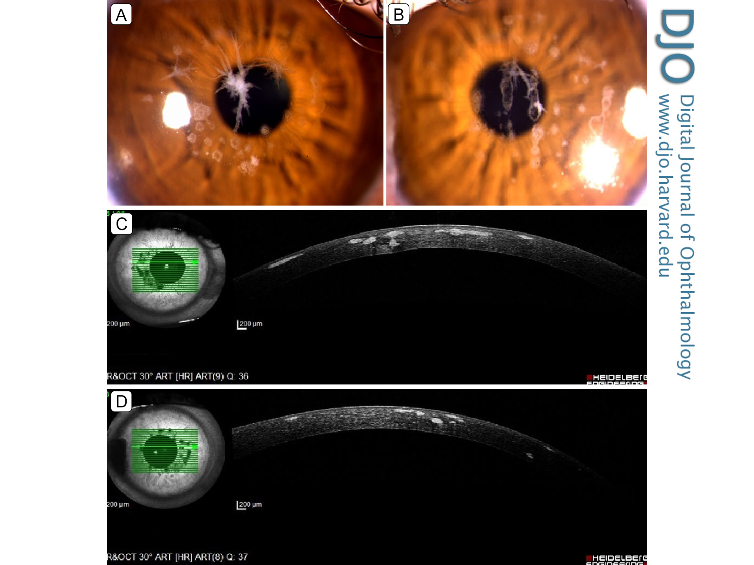
A 35-year-old woman presented at the Advanced Eye Center, Post Graduate Institute of Medical Education and Research, Chandigarh, with a 6-month history of glare and painless diminution of vision in both eyes. On examination, her best-corrected visual acuity was 20/20 in the right eye and 20/60 in the left eye. White “bread crumb” opacities with “moth-eaten” centers were observed in the superficial and mid-corneal stroma (A, B). These were interspersed with star-shaped, spiny, or icicle-shaped deposits as seen in anterior segment optical coherence tomography (C, D). These features are consistent with Avellino dystrophy, also known as combined granular-lattice dystrophy, or granular corneal dystrophy, type 2 (GCD2). It is caused by a mutation in the TGFBI (transforming growth factor beta-induced) gene and is characterized by both amyloid and hyaline deposits in the corneal stroma. Patients are treated with phototherapeutic keratectomy or with penetrating keratoplasty. Because our patient had good vision, only spectacle correction was advised, and she continued to be followed for disease progression.
 Welcome, please sign in