|
|
|
Register
with DJO to receive personalized updates.
If you're already a
member, please sign in.
|
|
|
|
|
|
|
|
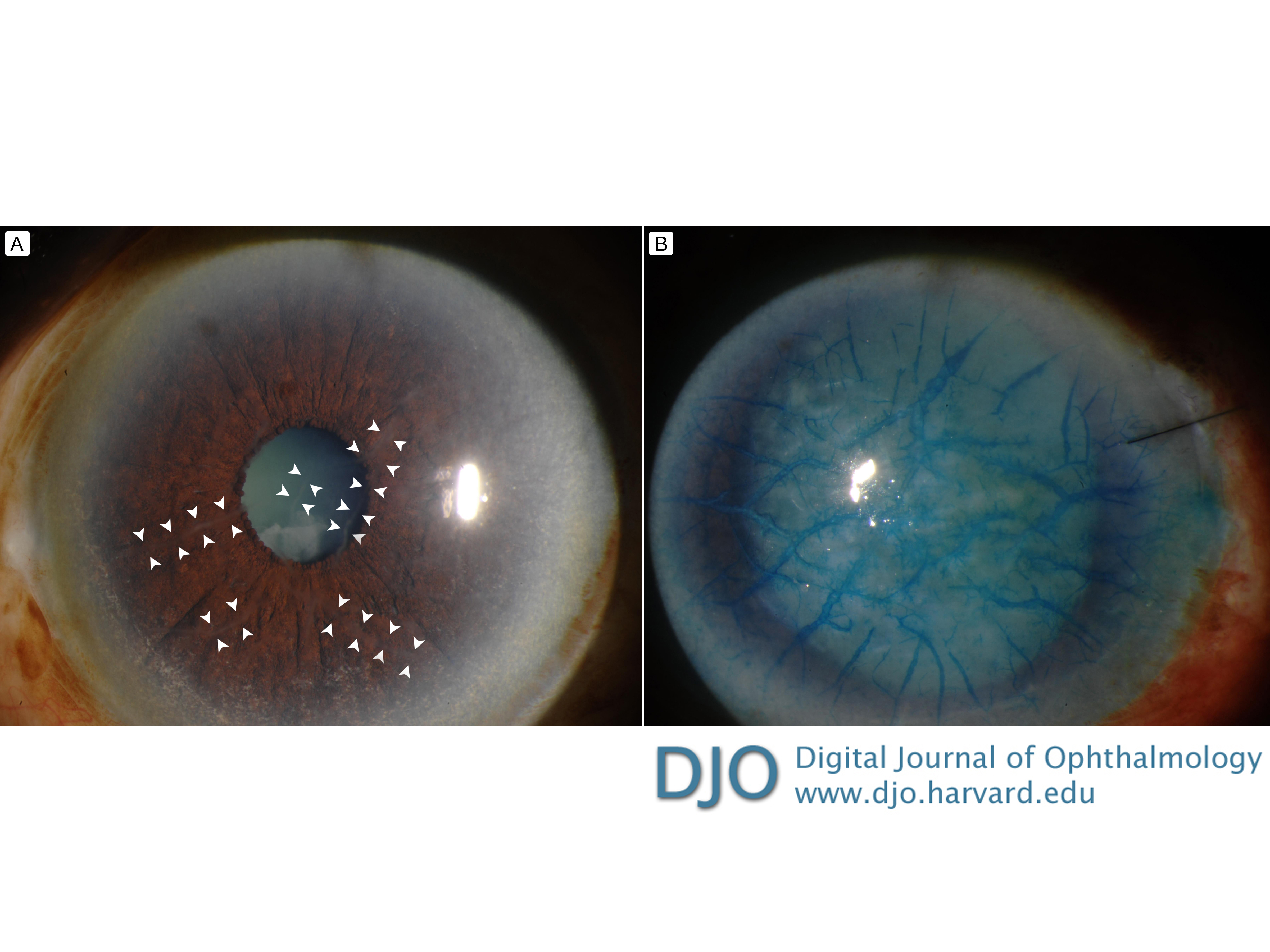
Lattice corneal dystrophy Aug 26, 2019 Volume 25, Number 3 Osama M. Mustafa, MD | Cornea Division, Wilmer Eye Institute, Johns Hopkins University School of Medicine, Baltimore, Maryland Yassine J. Daoud, MD, FACS | Cornea Division, Wilmer Eye Institute, Johns Hopkins University School of Medicine, Baltimore, Maryland  A 77-year-old woman with a history of glaucoma presented at the Wilmer Eye Institute with decreased vision of several months’ duration. She had also experienced occasional episodes of redness, itchiness, and burning sensation prior to presentation. On examination, the best-corrected visual acuity was 20/80 in the right eye and 20/25 in the left eye. Anterior segment examination of the right eye revealed a cataract and subtle opaque streaks in the corneal stroma (A, arrowheads). The remainder of the examination was unremarkable. Cataract surgery was planned, but was complicated by severe intraocular pressure elevation refractory to anterior chamber taps and intravenous mannitol, resulting in corneal haziness. To help visualize the lens capsule intraoperatively, trypan blue was injected into the anterior chamber of the eye. With significant and persistent obscuration of the surgical view, the surgery was aborted. Postoperative examination showed peculiar staining of the radially oriented, branching, refractile, filamentous, subepithelial and stromal opacities by trypan blue (B). |
|
|
|
|
|
 Welcome, please sign in