
|
|
|
Register
with DJO to receive personalized updates.
If you're already a
member, please sign in.
|
|
|
|
|
|
|
|
Bilateral multifocal choroidal osteoma with choroidal neovascular membrane Jan 13, 2019 Volume 25, Number 1 Nagesha Krishnappa, MS | Department of Retina and Vitreous, Aravind Eye Care System, Pondicherry, India Pratyusha Ganne, MS | Department of Retina and Vitreous, Aravind Eye Care System, Pondicherry, India 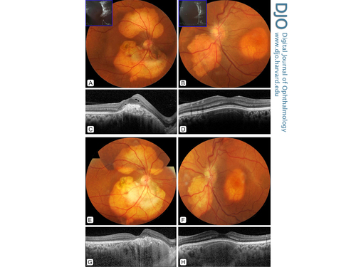 A healthy 32-year-old woman presented at Aravind Eye Hospital, Pondicherry, with gradual vision loss in her right eye (20/80) of 1 year’s duration. Fundus examination of the right eye showed two discrete yellowish-white subretinal lesions in the peripapillary area (A), pigmentary changes of the macula, and a dull foveal reflex. The fundus examination of the asymptomatic left eye showed a similar B-scan ultrasonography revealed hypereflectivity at the choroidal level that corresponded to the lesions, with back shadowing confirming the lesions’ calcific nature, suggestive of bilateral, multifocal choroidal osteoma (A-B, inset). Optical coherence tomography showed a choroidal neovascular membrane (CNVM), with intraretinal fluid in the right eye (C) and a dry macula in left eye (D). Investigations for secondary causes of the calcific deposits were negative, including calcium levels, thyroid profile, and blood counts. The patient received 2 doses of intravitreal bevacizumab. Following treatment, the CNVM remained inactive over 12 months’ follow-up (E and G). The left eye macula remained dry throughout the course of the disease (F and H). |
|
|
|
|
|
 Welcome, please sign in


