
|
|
|
Register
with DJO to receive personalized updates.
If you're already a
member, please sign in.
|
|
|
|
|
|
|
|
Laser pointer injury findings on OCT angiography Jan 1, 2019 Volume 25, Number 1 S. Tammy Hsu, BA | Duke University School of Medicine, Durham, North Carolina Lejla Vajzovic, MD | Duke University School of Medicine, Durham, North Carolina 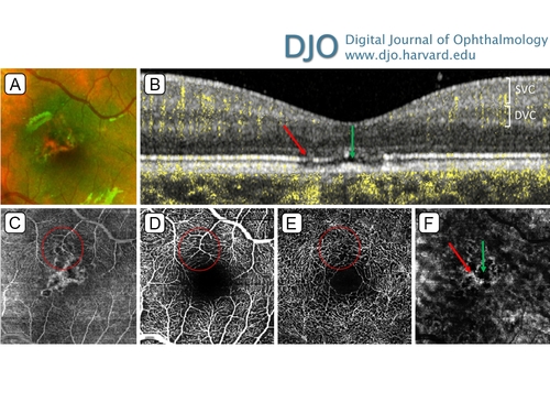 A healthy 6-year-old boy presented at the Duke Eye Center with decreased visual acuity to 20/30 and macular pigmentary changes in the left eye only (A). Optical coherence tomography (OCT) showed multifocal disruption of the foveal ellipsoid zone, with atrophy of the retinal pigment epithelium (RPE), pathognomonic for laser pointer-induced retinal injury (B). The parents confirmed the presence of laser pointers at home. The figure includes corresponding transverse OCT-angiography images: full A-scan compilation (C), superficial vascular complex (SVC; D), deep vascular complex (DVC; E), and the choroid (F). The injuries primarily localize to the foveal avascular zone, but there were possible loss of SVC and DVC capillaries overlapping with the areas of RPE disruption (example area circled). OCT-angiography of the choroid showed areas of hyper- and hypo-reflectivity; examination of the corresponding OCT B-scans showed that the choroidal shadowing was due to the hyperreflective mounds (B and F, green arrows) and that the choroidal enhancement was due to increased transmission through RPE defects (B and F, red arrows). Follow-up at 4 months showed no evidence of active disease process in either eye. |
|
|
|
|
|
 Welcome, please sign in


