
|
|
|
Register
with DJO to receive personalized updates.
If you're already a
member, please sign in.
|
|
|
|
|
|
|
|
Primary choroidal vitiligo Sep 15, 2018 Volume 24, Number 3 Manavi D. Sindal, MS, FMRF | Department of Vitreo-Retina, Aravind Eye Hospital, Puducherry, India Pratyusha Ganne, MS | Department of Vitreo-Retina, Aravind Eye Hospital, Puducherry, India 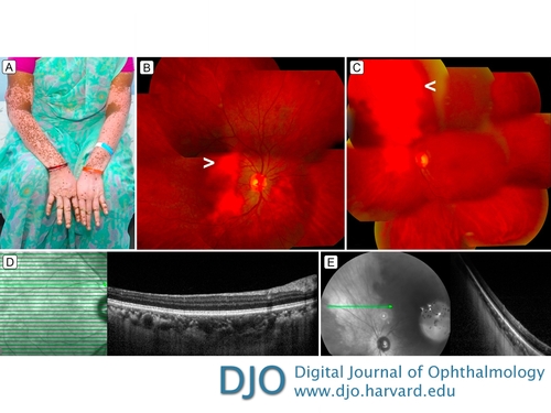 A 43-year-old woman with cutaneous vitiligo (A) presented at the Aravind Eye Hospital for a routine eye examination. Visual acuity was 20/20 in the right eye and 20/40 in the left eye. Anterior segment examination was normal in the right eye and showed an early-stage cataract in the left eye. There were no signs of ocular inflammation. Fundus examination showed a flat, depigmented patch in the macula of the right eye and superonasal to the optic disc in the left eye (B-C, arrowheads). Enhanced-depth optical coherence tomography (EDI-OCT) scans through the lesions showed normal retinal and choroidal layers (D-E). (The blur in E is an unavoidable artifact associated with use of a 50° lens with the Spectralis OCT [Heidelberg Engineering, Heidelberg, Germany]). Ultrasonography was normal. A diagnosis of choroidal vitiligo was made; our patient had no underlying autoimmune disease and no signs of previous ocular inflammation. The differential diagnosis for flat depigmented fundus lesions include amelanotic choroidal nevus, choroidal osteoma, diffuse choroidal hemangioma, amelanotic melanomas, and chorioretinal atrophy, all of which show characteristic abnormalities on EDI-OCT or ultrasonography. |
|
|
|
|
|
 Welcome, please sign in


