
|
|
|
Register
with DJO to receive personalized updates.
If you're already a
member, please sign in.
|
|
|
|
|
|
|
|
Hemorrhagic papilledema following lumbar puncture Nov 8, 2017 Volume 23, Number 4 Atalie C. Thompson, MD, MPH | Duke University, Department of ophthalmology, Durham, North Carolina Mays A. El-Dairi, MD | Duke University, Department of ophthalmology, Durham, North Carolina 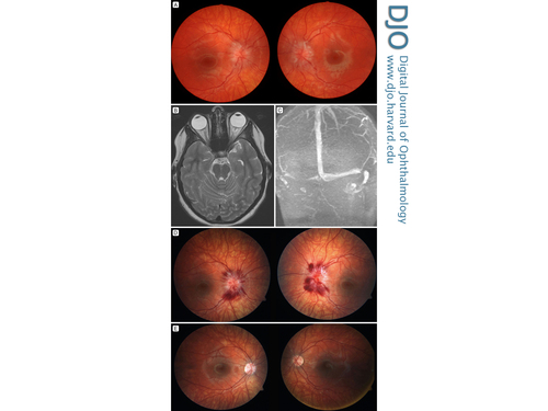 A 12-year-old prepubertal girl presented emergently to Duke Hospital with a 3-week history of headache, intracranial noises, and horizontal diplopia. On examination, visual acuity was 20/20 in both eyes. Humphrey visual fields were reliable, with mildly enlarged blind spots. Initial dilated fundus examination showed bilateral grade 4 papilledema on the Frisén scale (A). T2-weighted magnetic resonance imaging demonstrated posterior flattening of both globes, with mild prominence of the distal optic sheaths (B). Magnetic resonance venography showed right dominance of the transverse and jugular sinuses, with focal flattening of the dural venous sinuses at the junction of the transverse and sigmoid sinuses, typical of idiopathic intracranial hypertension (C). No mass or venous sinus thrombosis was identified. Lumbar puncture (LP) opening pressure was 36 cm H2O (closing pressure, 10 cm H2O) and cerebral spinal fluid labs were normal, consistent with pediatric intracranial hypertension. Post–lumbar puncture dilated fundus examination revealed interval development of 360° of peripapillary hemorrhages (D), although the optic nerves appeared less elevated. Hemorrhagic papilledema is classically associated with an increased risk of vision loss. However, our patient did not develop prominent disc hemorrhages until after undergoing large-volume LP. Given her normal visual function, the patient was treated medically with acetazolamide. Her papilledema and hemorrhages resolved over 3 weeks (E). We suspect that the substantial decrease in intracranial pressure during LP may have precipitated the swollen peripapillary vessels to rupture, leading to the development of hemorrhagic papilledema. |
|
|
|
|
|
 Welcome, please sign in


