
|
|
|
Register
with DJO to receive personalized updates.
If you're already a
member, please sign in.
|
|
|
|
|
|
|
|
Extraocular muscle belly enlargement with myotendinous sparing in thyroid-associated orbitopathy Dec 29, 2017 Volume 23, Number 4 Angela Gauthier, AB | Yale School of Medicine, New Haven, Connecticut Ichiro Ikuta, MD | Department of Neuroradiology, Yale School of Medicine, New Haven, Connecticut William B. Zucconi, DO | Department of Neuroradiology, Yale School of Medicine, New Haven, Connecticut 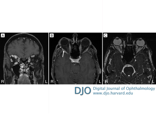 A 66-year-old man with Graves disease presented at the Yale New Haven Hospital for evaluation of worsening binocular diplopia. TSH levels were low (<0.005 μIU/mL), and thyroid peroxidase antibodies were elevated (555 IU/mL [normal, <35 IU/mL]). Coronal (A) and axial (B) T1-weighted magnetic resonance imaging with contrast showed bilateral enlargement of inferior, medial, superior, and lateral rectus muscle bellies (B, thick arrow), with sparing of the myotendinous junctions (B, thin arrow), consistent with thyroid-associated orbitopathy. Axial T2-weighted imaging (C) showed right globe anterior positioning measured from the interzygomatic line (normal, below 21 mm). Thyroid-associated orbitopathy affects 20%-50% of patients with Graves disease. It may be caused by autoantibody activation of orbital fibroblasts, causing inflammation and glycosaminoglycan deposition. The condition affects bilateral extraocular muscles 90% of the time; the inferior rectus muscle is most likely to be involved, followed by the medial rectus, superior rectus, lateral rectus, and oblique muscles. |
|
|
|
|
|
 Welcome, please sign in


