|
|
|
Register
with DJO to receive personalized updates.
If you're already a
member, please sign in.
|
|
|
|
|
|
|
|
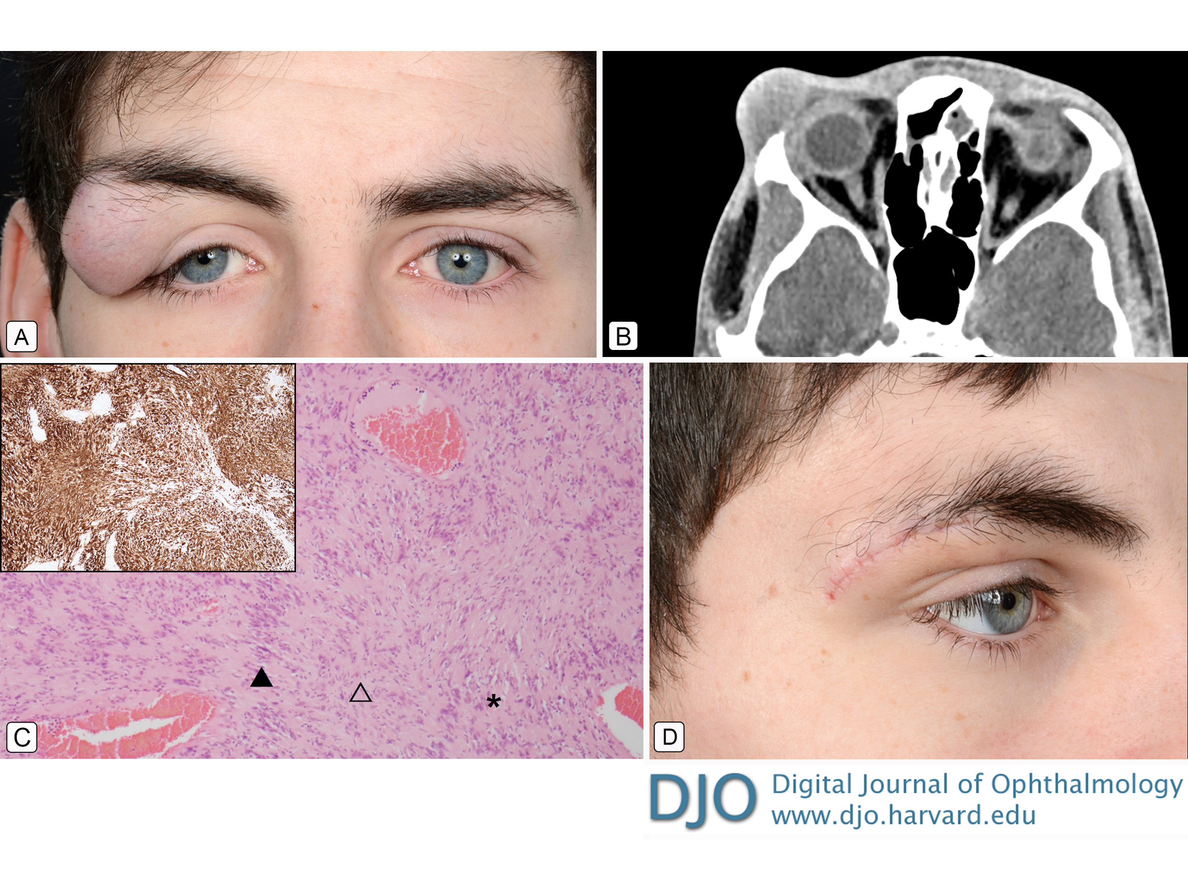
Giant eyelid schwannoma Jan 11, 2018 Volume 24, Number 1 Derek Kwun-hong Ho, MACantab | Department of Ophthalmology, Royal Gwent Hospital, Newport, UK Varsha Shah, FRCPath | Department of Pathology, Royal Gwent Hospital, Newport, UK Ebube E. Obi, FRCOphth | Department of Ophthalmology, Royal Gwent Hospital, Newport, UK  A 17-year-old young man presented at the Royal Gwent Hospital, United Kingdom, with a lump on his right upper eyelid that had been slowly increasing in size over the previous 5 years (A). Clinically it was suggestive of a dermoid cyst. Computed tomography revealed nonspecific, isolated soft tissue swelling on the lateral aspect of the right orbit, with no localized bone destruction (B). There was no evidence of neurofibromatosis. An excisional biopsy was performed, and histopathological examination (C) revealed a benign schwannoma measuring at 32 × 25 × 20 mm; the tumor was well circumscribed, with hypercellular Antoni A areas (empty triangle) and hypocellular Antoni B areas (asterisk) and interspersed dilated blood vessels. It was composed of spindle cells with wavy nuclei, arranged in palisades (Verocay bodies, filled triangle). No significant nuclear atypia or mitotic activity was observed. S-100 stain was diffusely positive, confirming the diagnosis (C, inset). The patient made a full recovery (D). |
|
|
|
|
|
 Welcome, please sign in