Lobulated uveal melanoma with both pigmented and nonpigmented tumor
Jan 28, 2018
Volume 24, Number 1
Linda A. Cernichiaro-Espinosa, MD | Retina and Vitreous, Asociación para Evitar la Ceguera en México IAP, México City, México
Sara González-Godínez, MD | Echography Department, Asociación para Evitar la Ceguera en México IAP, México City, México
Dolores Ríos y Valles-Valles, MD | Ophthalmic Pathology Service, Asociación para Evitar la Ceguera en México IAP, México City, México
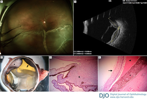
A 56-year-old man presented at the Asociación para Evitar la Ceguera en México with an inferior, exudative retinal detachment secondary to a uveal melanoma (A). Echography revealed a bilobulated choroidal solid tumor with medium-to-high reflectivity and retinal detachment (B). Enucleation was performed. The macroscopic appearance showed an amelanotic neoplasm in the ciliary body (asterisk) and a pigmented tumor (arrow) in the choroid with the overlying retina detached (C). Histopathologic examination (D,E [hematoxylin-eosin, original magnification ×200]) revealed an amelanotic tumor with widening of the ciliary body at the pars plicata (D, asterisk) and a pigmented neoplasm (E, asterisk) with the associated exudative (E, arrowhead) detachment of the retina (E, arrow).
 Welcome, please sign in


