Vortex vein varix simulating choroidal melanoma
Nov 28, 2016
Volume 22, Number 4
Maria Pefkianaki, MD, PhD | Ocular Oncology Service, Wills Eye Hospital,Thomas Jefferson University, Philadelphia, Pennsylvania
Timothy S. Fuller, MD | Ocular Oncology Service, Wills Eye Hospital,Thomas Jefferson University, Philadelphia, Pennsylvania
Carol L. Shields, MD | Ocular Oncology Service, Wills Eye Hospital,Thomas Jefferson University, Philadelphia, Pennsylvania
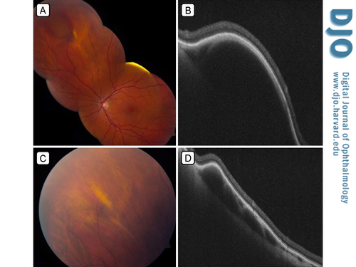
A 46-year-old, white woman presented at the Ocular Oncology Clinic of Wills Eye Hospital with a dome-shaped, dark, elevated mass in the superonasal equatorial region of the left eye. The lesion was prominent on superonasal gaze and flattened on application of digital pressure to the globe (A,C). Spectral domain optical coherence tomography demonstrated an optically lucent choroidal mass (B), which flattened when light digital pressure was applied to the globe (D). The vortex veins, typically located at the equator of the globe, allow for egress of choroidal blood from the eye. The confluence of choroidal vessels drain into an ampulla, which drains into the vortex vein. Dilation of the ampulla is known as a varix. A varix has gaze-related dynamic characteristics, with vascular filling and draining. Varices can be clinically confused with a choroidal melanoma, choroidal nevus, or subretinal hemorrhage; confusion with these entities, however, can lead to extensive evaluation and cause unnecessary patient stress.
 Welcome, please sign in


