
|
|
|
Register
with DJO to receive personalized updates.
If you're already a
member, please sign in.
|
|
|
|
|
|
|
|
Uveal melanoma with BAP1 mutation Jun 23, 2016 Volume 22, Number 2 Jin Soo Andy Song, BSc | Dalhousie University Adam Dmytriw, MD, MSc | University of Toronto Hesham Lakosha, MD | Dalhousie University 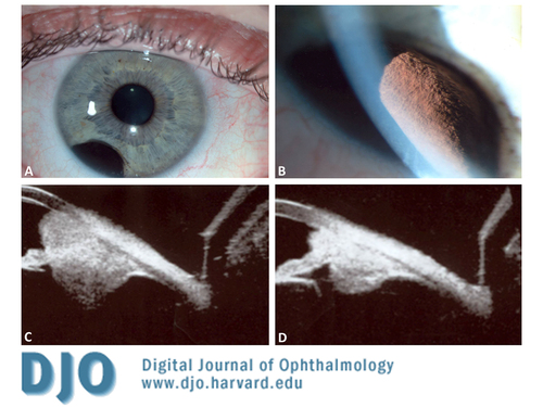 A 40-year-old, healthy man presented with a darkly pigmented inferior temporal lesion in his right eye for 1 year. The corrected visual acuity, intraocular pressure, and neurological examination were unremarkable. Slit-lamp biomicroscopy showed a demarcated pigmented lesion in the peripheral iris between 6 o'clock and 8 o'clock (A). The estimated basal diameter for this mass measured 4mm x 2.5 mm (B). Ultrasound biomicroscopy (UBM) revealed a mass deriving from the iris stroma without ciliary body involvement, suggesting an iris origin (C). Fine-needle aspiration (FNA) confirmed melanoma with inactivation of the BAP1 gene, increasing the probability of metastasis and prompting treatment. UBM has become crucial in differentiation of uveal melanomas from benign growths, as lesions <3 mm cannot be reliably visualized by other imaging modalities. The patient was treated with brachytherapy using an I-125 plaque. Follow-up UBM 3 years later illustrated successful radiotherapy with a reduced tumor (D). |
|
|
|
|
|
 Welcome, please sign in


