|
|
 |
|
|
|
|
|
A 57-year-old man with leukocytosis and sphenoid sinus disease
Digital Journal of Ophthalmology 2020
Volume 26, Number 2
April 24, 2020
DOI: 10.5693/djo.03.2019.09.003
|
Printer Friendly
|
|
|

Ansuya P. Deosaran, MD | Department of Ophthalmology, Louisiana State University, New Orleans, Louisiana Ahmaida Zeglam, MD | Department of Ophthalmology, University of Florida, Gainesville, Florida Mary K. Wilson, BS | College of Medicine, University of Florida, Gainesville, Florida Andres Gonzalez, MD | Department of Ophthalmology, University of Florida, Gainesville, Florida Matthew J. Gray, MD | Department of Ophthalmology, University of Florida, Gainesville, Florida
|
|
|
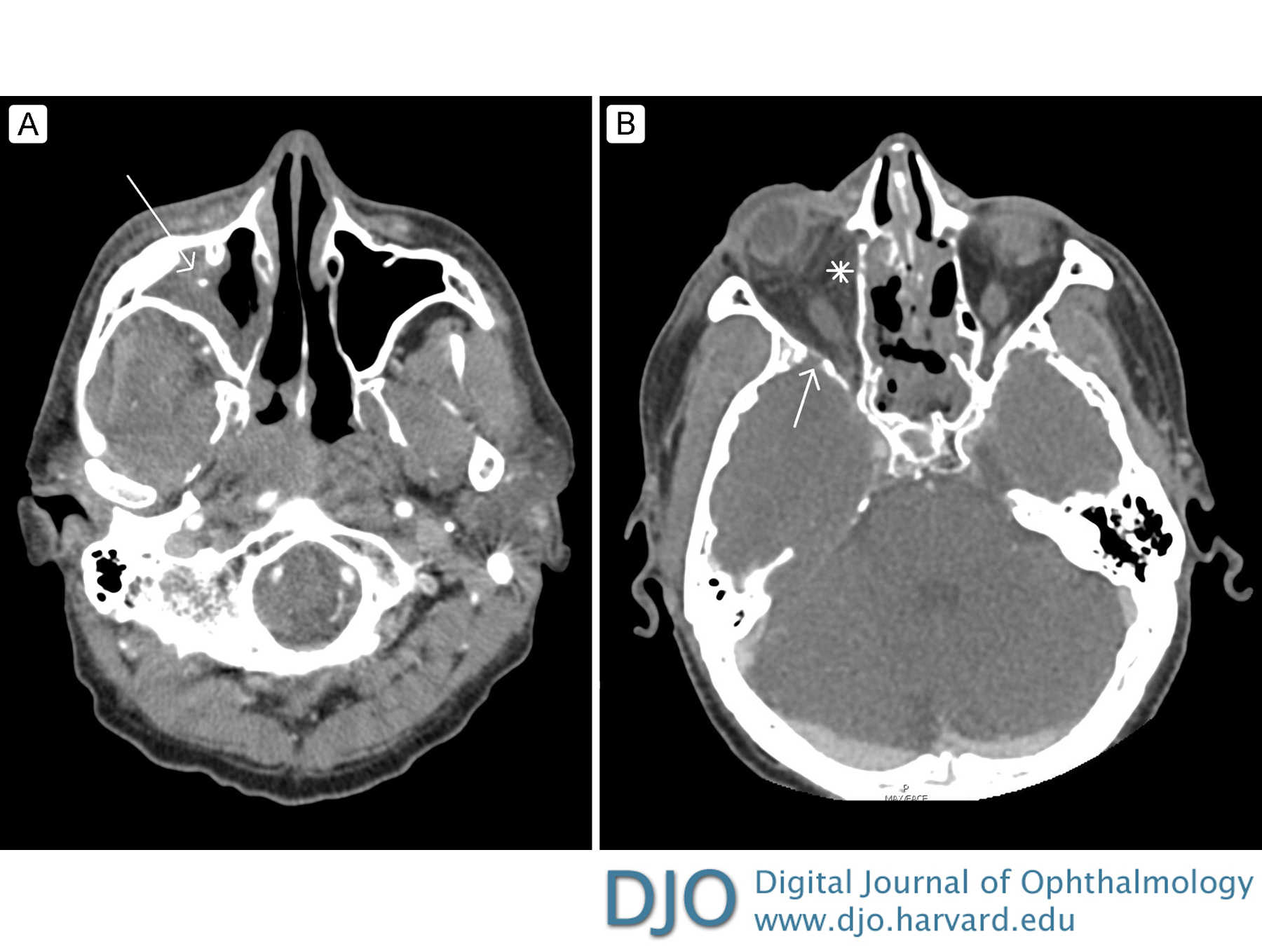
| History | | A 57-year-old man with a past medical history of coronary artery disease, chronic sinusitis, allergic rhinitis, and history of sinus surgery on intranasal steroids presented to an outside emergency room with worsening sinusitis symptoms of 4-5 days’ duration. On arrival, labs revealed a leukocytosis, and computed tomography (CT) of the maxillofacial region revealed significant sphenoid sinus disease, with mild mucosal thickening in the right ethmoid and frontal sinuses. He was admitted for treatment with antibiotics. The following day, the patient felt his swelling and pain had improved but noted acutely decreased vision in his right eye. CT angiography of the head on this day revealed right periorbital edema and cellulitis, with occlusion versus thrombus of the right superior ophthalmic vein and engorgement of the medial and inferior rectus muscles. There was also opacification of the right sphenoid and maxillary sinus (Figure 1). He was started on heparin by Neurology. Two days after admission, he experienced increasing right periorbital swelling, redness, and ptosis. He was transferred to University of Florida, Gainesville, for ophthalmological examination. | |
|
Figure 1
Thin slice, contrast-enhanced computed tomography (CT) in an axial plane at two levels show opacification of the right sphenoid and maxillary sinuses, indicated with arrows. Figure 1A demonstrates significant proptosis of the right eye, with early postseptal infiltration in the form of inflammatory stranding of the retro-orbital fatty tissue with reactive osteitis. Figure 1B, a lower axial cut, better demonstrates the opacification of the right maxillary sinus.
 |
|
|
|
|
|
|
|
 Welcome, please sign in