|
|
 |
|
|
|
|
|
A 71-year-old man with bilateral vision loss
Digital Journal of Ophthalmology 2017
Volume 23, Number 3
September 3, 2017
DOI: 10.5693/djo.03.2017.03.002
|
Printer Friendly
Download PDF |
|
|

Katy C. Liu, MD, PhD | Department of Ophthalmology, Duke University, Durham, North Carolina Jullia A. Rosdahl, MD, PhD | Department of Ophthalmology, Duke University, Durham, North Carolina Mays El-Dairi, MD | Department of Ophthalmology, Duke University, Durham, North Carolina
|
|
|
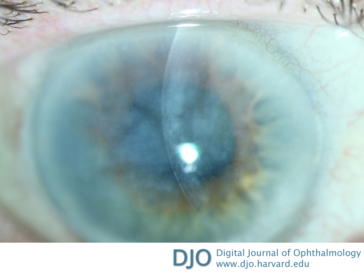
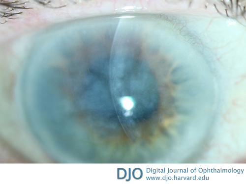
| Examination | Visual acuity in each eye was no light perception, without saccades to optokinetic drum. Pupils were fixed and dilated (on atropine). Intraocular pressure was 4 mm Hg in the right eye and 5 mmHg in the left eye. The external examination was notable for right upper eyelid ptosis. The slit-lamp examination of the right eye showed significant opacification and edema of the right cornea, no keratic precipitates, and a hazy view to the anterior chamber without frank hypopyon or posterior synechiae (Figure 1). Slit-lamp examination of the left eye showed trace corneal edema, no keratic precipitates, and no cell or flare in the anterior chamber.
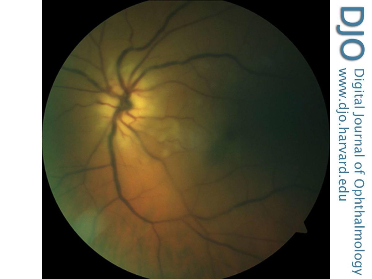
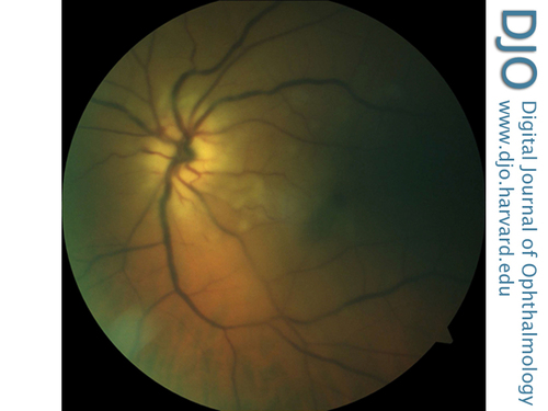
Dilated fundus examination of the left eye was limited but showed 3+ optic nerve head edema. There was a cherry red spot. Arterioles were attenuated, and venules were dark and severely dilated (Figure 2). The peripheral retina was attached. There was no view posteriorly in the right eye.
| |
|
Figure 1
Anterior segment photograph of the right eye demonstrating an edematous and opacified cornea without keratic precipitates.
 |
|
|
Figure 2
Fundus photograph of the left eye demonstrating a cherry-red spot, attenuated arterioles, and dark, dilated venules.
 |
|
|
|
|
|
|
|
 Welcome, please sign in