|
|
 |
|
|
|
|
|
A 71-year-old man with bilateral vision loss
Digital Journal of Ophthalmology 2017
Volume 23, Number 3
September 3, 2017
DOI: 10.5693/djo.03.2017.03.002
|
Printer Friendly
Download PDF |
|
|

Katy C. Liu, MD, PhD | Department of Ophthalmology, Duke University, Durham, North Carolina Jullia A. Rosdahl, MD, PhD | Department of Ophthalmology, Duke University, Durham, North Carolina Mays El-Dairi, MD | Department of Ophthalmology, Duke University, Durham, North Carolina
|
|
|
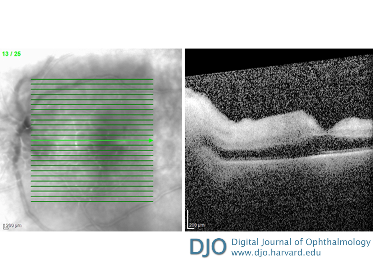
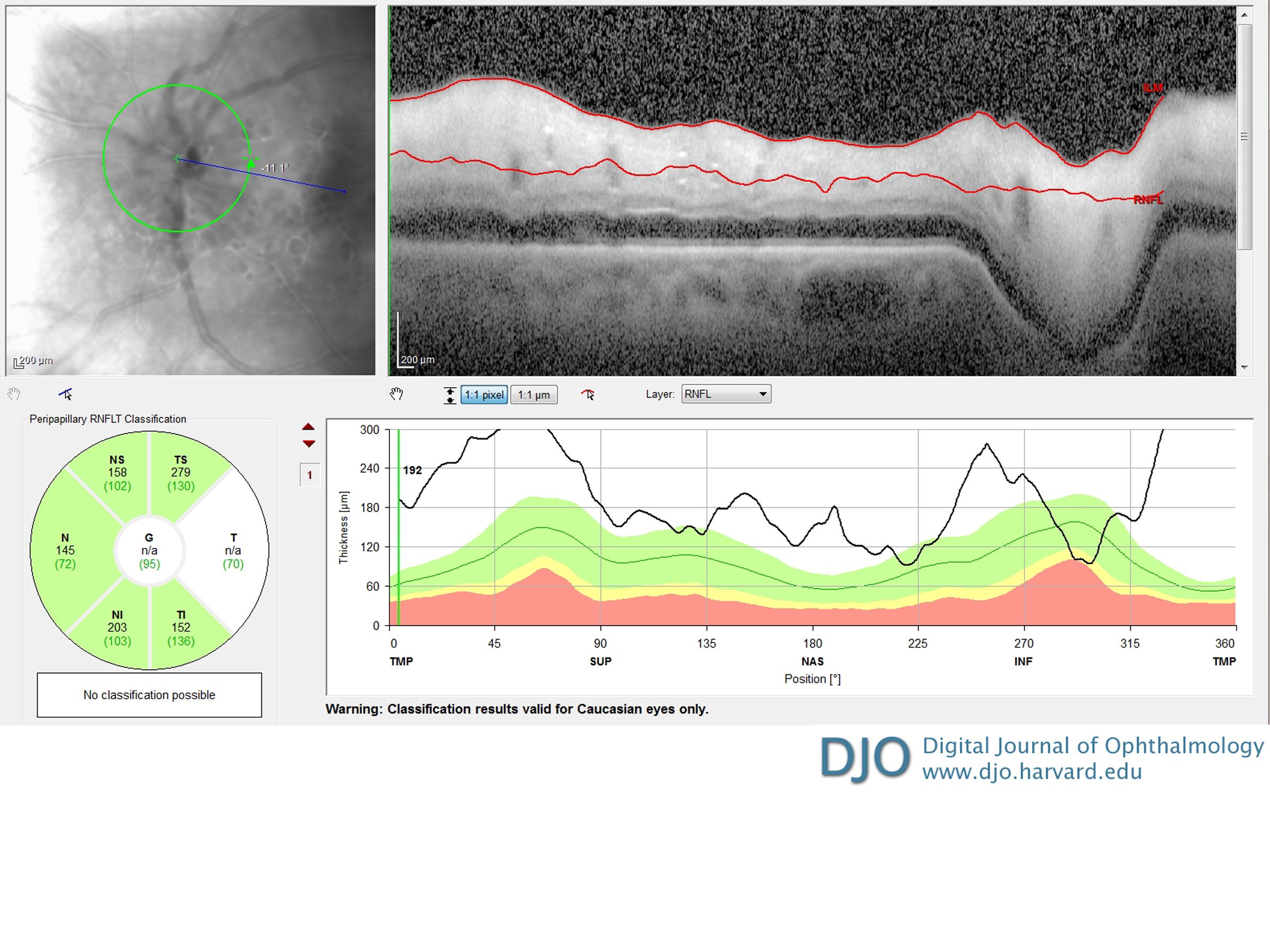
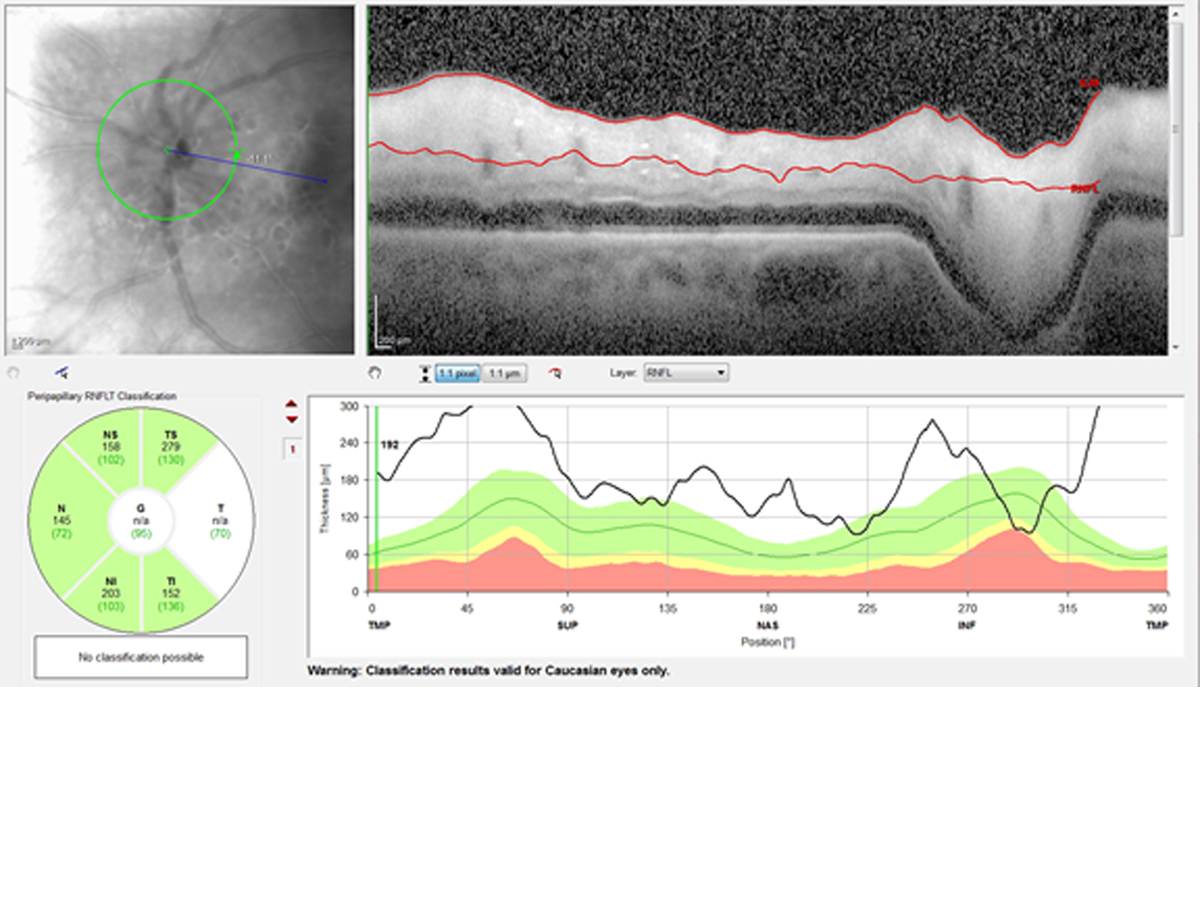
| Ancillary Testing | B-scan ultrasonography of the right eye showed no vitritis, posterior mass, or retinal detachment. In the left eye, optical coherence tomography (OCT) of the macula revealed diffuse macular thickening and enlarged venules (Figure 3). OCT of the retinal nerve fiber layer (RNFL) showed optic nerve head edema (Figure 4).
Complete blood count was within normal limits with the exception of an elevated platelet count (484 ×103/mm3). Basic metabolic panel was remarkable for low sodium (132 mEq/L), potassium (2.3 mEq/L), chloride (95 mEq/L), and brain natriuretic peptide (6 mg/dL). Inflammatory markers were elevated: erythrocyte sedimentation rate (ESR) was 59 mm/hr, and C-reactive protein (CRP) was 7.53 mg/L (normal range, 0.02-0.7 mg/L). Additional testing that returned negative after diagnosis included purified protein derivative skin test, rapid plasma reagin, fluorescent treponemal antibody absorption test, anti-neutrophil cytoplasmic antibody (ANCA), and antinuclear antibody tests.
Maxillofacial computed tomography (CT) demonstrated no abnormalities. Magnetic resonance imaging (MRI) of the brain with and without contrast showed mild atrophy and chronic microvascular ischemic changes. | |
|
Figure 3
Optical coherence tomography (OCT) of the left eye revealed a diffusely thickened macula without fluid.
 |
|
|
Figure 4
OCT retinal nerve fiber layer of the left eye showing thickening of the retinal nerve fiber layer around the disc suggesting optic nerve head edema.
 |
|
|
|
|
|
|
|
 Welcome, please sign in