|
|
 |
|
|
|
|
|
A 35-year-old woman presenting with pain, reduced right-eye vision, and headache
Digital Journal of Ophthalmology 2015
Volume 21, Number 2
May 13, 2015
DOI: 10.5693/djo.03.2015.03.002
|
Printer Friendly
Download PDF |
|
|

Brinda P. Shah, MS, FRCOphth | Moorfields Eye Hospital, London, United Kingdom Jonathan Clarke, MD, FRCOphth | NIHR Biomedical Research Centre at Moorfields Eye Hosital NHS Foundation Trust and UCL Institute of Ophthalmology, London, United Kingdom
|
|
|
| Diagnosis and Discussion | Pediatric cataract surgery is still often performed without the insertion of an intraocular lens. Hitchings described acute, aphakic, pupillary-block glaucoma as “a relatively uncommon condition characterized by ocular hypertension, shallowing of the anterior chamber and marked convexity of the iris, together with angle closure.”(1) The pupil closes onto the vitreous, obstructing the route for aqueous to egress from the posterior chamber to the anterior chamber. Positive pressure in the posterior chamber in comparison to the anterior chamber forces vitreous anteriorly. The iris is also pushed anteriorly by the positive posterior chamber pressure. This explains the shallow anterior chamber and secondary closure of the iridocorneal angle. It can occur with an intact or broken anterior hyaloid face, but in both instances, vitreoendothelial contact is the rule.(1)
In this situation, Shaffer also describes the role of miotics in increasing the tension of the pupil causing a plugging effect on the vitreous thereby creating a resistance to aqueous outflow.(2) This may explain the reason for sunlight acting as a trigger for headaches in our patient, which were likely intermittent episodes of pupillary block.
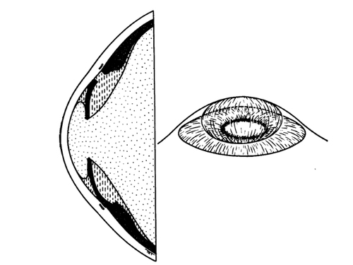
An illustration from Hitching’s 1979 paper is remarkably similar to the findings documented on the day of the acute episode (Figure 2).(1) There have been several reports of intermittent-angle closure episodes (in phakic but not aphakic patients) misdiagnosed as migraine prior to arriving at the correct diagnosis. In these cases, there was an association with high hypermetropia or shallow anterior chambers, prompting ophthalmology referral. Assessment for angle width confirmed intermittent-angle closure.(3-5)
In aphakic pupillary block, the anterior chambers can be deep, except in an acute setting. Posner has described 3 stages of pupillary block by the vitreous, as follows: (1) an early stage, characterized by relative block due to decreased permeability of the hyaloid face; (2) a moderate stage, with adhesions between the vitreous and sphincter pupillae referred to as sphincteric pupillary block; and (3) the irido-hyloidal stage, with extensive adhesions between the anterior hyaloid face and the entire posterior iris surface.(6)
The diagnosis requires careful history taking and identification of precipitating factors, such as those leading to pupil constriction. These factors include bright sunlight (as in the present case) or parasympathomimetics such as pilocarpine. The presence of vitreous at the pupil axis should raise concern about the possibility of future pupil block. Pupil block glaucoma is very unusual in the presence of a patent iridectomy.
The patient was managed with medical treatment, including topical atropine until the definitive surgical treatment. A laser iridotomy might also have broken the pupil block, but the iris can be edematous with high IOP preventing a straightforward laser iridotomy.
A change in the nature of symptoms should alert the treating physician to look for pathology other than migraine to explain the nature of the symptoms.
Acknowledgments
Supported by the National Institute for Health Research (NIHR) Biomedical Research Centre based at Moorfields Eye Hospital NHS Foundation Trust and UCL Institute of Ophthalmology. The views expressed are those of the authors and not necessarily those of the NHS, the NIHR or the Department of Health. | |
|
Figure 2
Cross-section and diagrammatic three-dimensional view indicating relationships between vitreous, aqueous and iris in aphakic pupil block.1 (Reproduced with permission from Hitchings RA. Acute aphakic pupil block glaucoma: an alternative surgical approach. Br J Ophthalmol 1979;63:31-7.)
 |
|
|
|
|
|
|
|
 Welcome, please sign in


