|
|
 |
|
|
|
|
|
A 35-year-old woman presenting with pain, reduced right-eye vision, and headache
Digital Journal of Ophthalmology 2015
Volume 21, Number 2
May 13, 2015
DOI: 10.5693/djo.03.2015.03.002
|
Printer Friendly
Download PDF |
|
|

Brinda P. Shah, MS, FRCOphth | Moorfields Eye Hospital, London, United Kingdom Jonathan Clarke, MD, FRCOphth | NIHR Biomedical Research Centre at Moorfields Eye Hosital NHS Foundation Trust and UCL Institute of Ophthalmology, London, United Kingdom
|
|
|
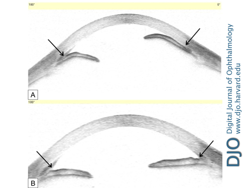
| Ancillary Testing | | Anterior segment optical coherence topography was performed and demonstrated pupillary-block glaucoma, with anterior displacement of the iris and closure of the iridocorneal angles (Figure 1A). | |
|
Figure 1
A, Anterior segment optical coherence topography of patient with pupillary-block glaucoma with elevated intraocular pressure showing anterior bowing of the iris and closure of the iridocorneal angle (arrows). B, After treatment, the anterior chamber has deepened with open angles and a flat iris contour (arrows).
 |
|
|
|
|
|
 Welcome, please sign in


