|
|
 |
|
|
|
|
|
A 71-year-old woman with decreased vision, nyctalopia, and peripheral vision loss
Digital Journal of Ophthalmology 2016
Volume 22, Number 4
December 31, 2016
DOI: 10.5693/djo.02.2016.06.001
|
Printer Friendly
Download PDF |
|
|
 Ravi Parikh, MD, MPH
 | Department of Ophthalmology and Visual Science, Yale University, New Haven, Connecticut Miguel A. Materin, MD | Department of Ophthalmology and Visual Science, Yale University, New Haven, Connecticut; Smilow Hospital at Yale New Haven Hospital Robert Lesser MD | Eye Care Group, New Haven, Connecticut Joachim Baehring, MD, PhD | Smilow Hospital at Yale New Haven Hospital; Department of Neurology, Yale University, New Haven, Connecticut Mario Sznol, MD | Smilow Hospital at Yale New Haven Hospital; Department of Internal Medicine, Yale University, New Haven, Connecticut Jennifer A. Galvin MD | Department of Ophthalmology and Visual Science, Yale University, New Haven, Connecticut
|
|
|
| Examination | On initial presentation, best-corrected visual acuity measured 20/20+ in each eye. Three months later, on presentation at Yale, visual acuities were 20/25 +2 in the right eye and at 20/30 −1 in the left eye. Anterior segment examination in each eye was unremarkable except for 1+ nuclear sclerotic cataract in the right eye and trace cortical cataract in the left eye. Pupils were round and equally reactive, and there was no relative afferent pupillary defect. Ishihara plates were 14/14 in each eye. Ocular motility was full, and the patient was orthotropic.
Dilated fundus examination of the right eye showed no vitreous cells, a titled, sharp, pink optic nerve, and a flat macula with a good foveal reflex. In the left eye, there were 2+ vitreous cells with a tilted optic nerve with temporal pallor and a trace epiretinal membrane in the macula. Both optic nerves had a cup-to-disc ratio of 0.1. Retinal vasculature in both eyes showed mild arteriolar attenuation.
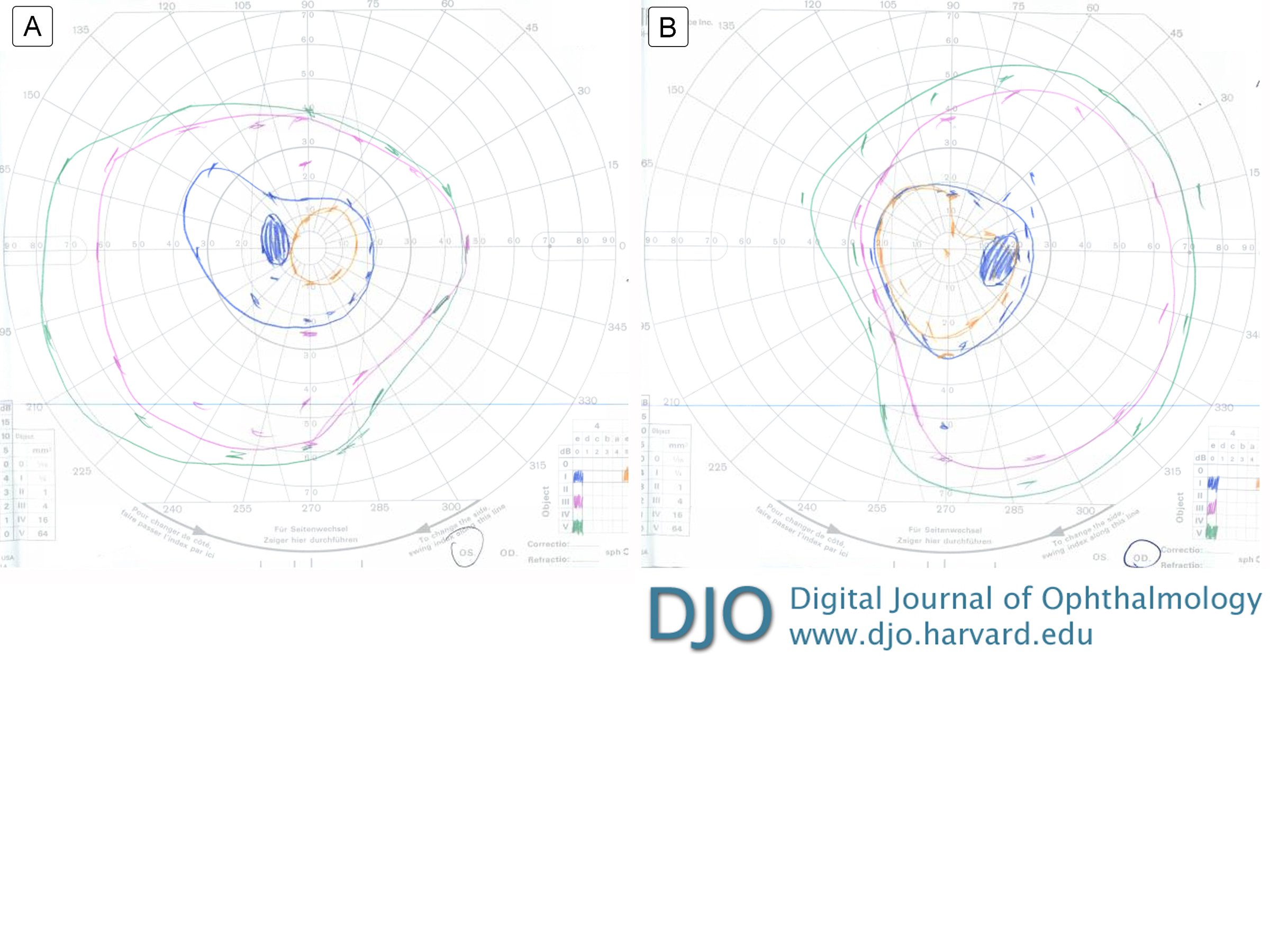
Goldmann visual field testing of the right eye showed I-4e reduced to 20° circumferentially and V-4e with mild reduction superiorly to 50° and mild reduction temporally to 70° (Figure 1A); in the left eye, I-3e reduced to 10° circumferentially and III-4e and V-4e moderately reduced to 35°-40° superiorly and reduced inferiorly to 55°-60° (Figure 1B). | |
|
Figure 1
Goldmann visual field: left eye (A), right eye (B).
 |
|
|
|
|
|
|
|
 Welcome, please sign in