|
|
 |
|
|
|
|
|
A 26-year-old man with a blind spot in his left eye
Digital Journal of Ophthalmology 2013
Volume 19, Number 3
September 26, 2013
DOI: 10.5693/djo.03.2013.07.001
|
Printer Friendly
Download PDF |
|
|

Alfred White Jr, MD | USF Eye Institute, University of South Florida Timothy Saunders, MD | USF Eye Institute, University of South Florida Peter Reed Pavan, MD | USF Eye Institute, University of South Florida
|
|
|
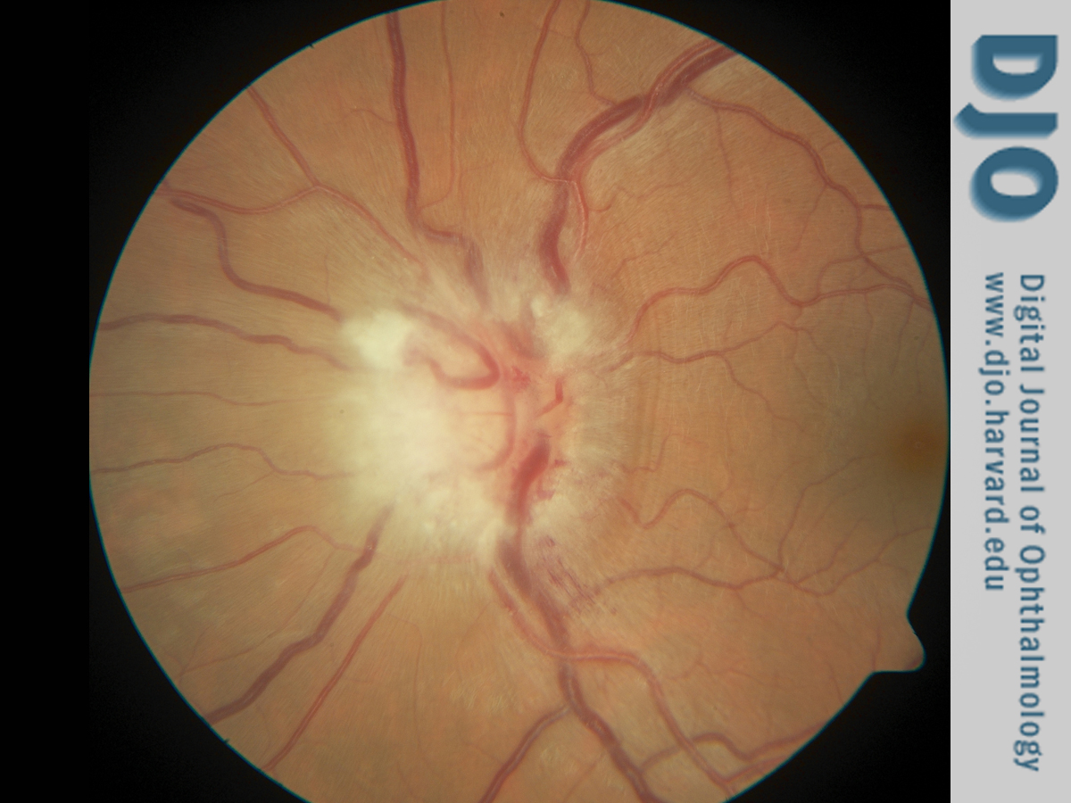
| Examination | Visual acuity was 20/20 in each eye, with intraocular pressures of 15 mm Hg in the right eye and 16 mm Hg in the left eye. Pupils were regular, round, and reactive. A relative afferent pupillary defect was present in the left eye. Extraocular movements were full in both eyes. The patient endorsed temporal visual field loss in the left eye on confrontation. Slit-lamp examination of the anterior segment was unremarkable.
Dilated examination of the right eye was within normal limits. Examination of the left eye was pertinent for grade 1 vitreous haze (Figure 1) in the photographic scale described by Davis.(1) There was moderate optic nerve edema with adjacent flame hemorrhages (Figure 1). A small, 1/2 disc diameter area of nonspecific chorioretinal changes was noted in the inferotemporal midperiphery without associated retinochoroiditis. | |
|
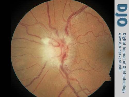
Figure 1
Color fundus photograph of the left eye. There is optic nerve edema and flame hemorrhage; vitreous haze, grade 1, was also appreciated.
 |
|
|
|
|
|
|
|
 Welcome, please sign in