|
|
 |
|
|
|
|
|
A 26-year-old man with a blind spot in his left eye
Digital Journal of Ophthalmology 2013
Volume 19, Number 3
September 26, 2013
DOI: 10.5693/djo.03.2013.07.001
|
Printer Friendly
Download PDF |
|
|

Alfred White Jr, MD | USF Eye Institute, University of South Florida Timothy Saunders, MD | USF Eye Institute, University of South Florida Peter Reed Pavan, MD | USF Eye Institute, University of South Florida
|
|
|
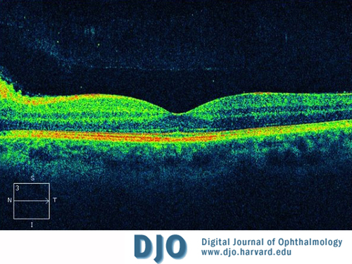
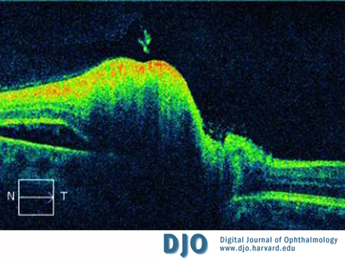
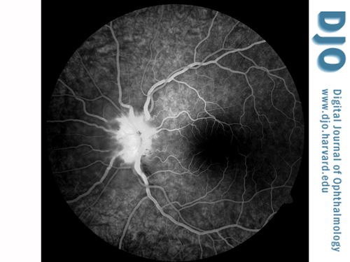
| Ancillary Testing | Optical coherence tomography (OCT) of the left eye showed peripapillary nerve fiber layer edema (Figure 2) and a small, shallow, serous retinal detachment nasal to the nerve (Figure 3). Late phase fluorescein angiography demonstrated staining of the left optic nerve (Figure 4). The macula displayed normal fluorescence.
The workup of this patient included complete blood count, basic metabolic panel, B12, folate, angiotensin converting enzyme (ACE), antinuclear antibody (ANA), Bartonella and toxoplasma serologies, and fluorescent treponemal antibody (FTA-Abs). HIV testing was deferred. A lumbar puncture was performed, with an opening pressure of 20 cm H2O. Cerebrospinal fluid was analyzed for cryptococcal antigen, oligoclonal bands, Herpes simplex virus (HSV) 1 and 2 polymerase chain reaction (PCR), cytomegalovirus (CMV) PCR, and India ink.
Magnetic resonance imaging (MRI) and magnetic resonance angiography (MRA) of the brain and orbits was performed with and without contrast, demonstrating normal signal of the optic nerves.
All of the test results were negative, except for Toxoplasma serologies, which were equivocal for Immunoglobulin M (IgM). This prompted repeat testing, which was positive for both IgG and IgM. | |
|
Figure 2
Macular optical coherence tomography (OCT) of the left eye showing peripapillary nerve fiber layer edema.
 |
|
|
Figure 3
Peripapillary OCT of the left eye. Nerve fiber layer thickening and a neurosensory detachment nasal to the nerve are present.
 |
|
|
Figure 4
Fluorescein angiography of the left eye at 1:05.7 demonstrating hyperfluorescence and staining of the optic nerve.
 |
|
|
|
|
|
|
|
 Welcome, please sign in


