|
|
 |
|
|
|
|
|
A 54-year-old woman with bluish discoloration of her sclera
Digital Journal of Ophthalmology 2010
Volume 16, Number 2
May 8, 2010
DOI: 10.5693/djo.03.2010.02.002
|
Printer Friendly
Download PDF |
|
|
 Isabella Phan, MD
 | Oregon Health and Science University Rachel Kaiser, MD, MPH | University of California, San Francisco Cynthia Chiu, MD | University of California, San Francisco
|
|
|
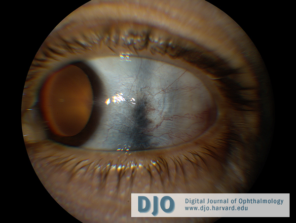
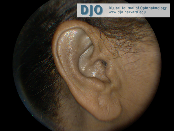
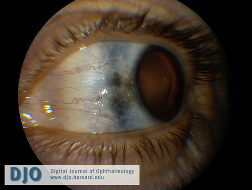
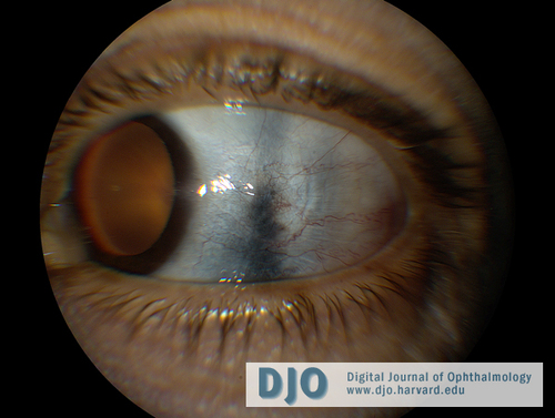
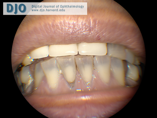
| Examination | | On examination, the patient’s visual acuity was 20/25 bilaterally. Her pupillary examination, intraocular pressures, extraocular movements, and confrontational visual fields were all normal. She had dark-brown and blue discoloration of the sclera circumferentially approximately 4 mm posterior to the limbus bilaterally, most concentrated nasally and temporally (Figures 1 and 2). There was no scleral thinning or injection. Her corneas were clear with no keratic precipitates. The anterior chambers were quiet. Her lenses, vitreous, and retinae were unremarkable, with no evidence of vasculitis, uveal tumors, or intraocular inflammation. Closer inspection of the patient's face revealed bluish discoloration of the lateral canthi and lower eyelid skin bilaterally, the pinna of both ears, and her front teeth (Figures 3 and 4). Her fingernails were unremarkable. There were no other areas of blue discoloration. | |
|
Figure 1
Left eye with bluish-gray scleral hyperpigmentation nasally.
 |
|
|
Figure 2
Left eye with bluish-gray scleral hyperpigmentation temporally.
 |
|
|
Figure 3
Bluish discoloration of the patient’s left ear.
 |
|
|
Figure 4
Bluish discoloration of the patient’s teeth and gums.
 |
|
|
|
|
|
|
|
 Welcome, please sign in