|
|
 |
|
|
|
|
|
Pupillary capture of implantable collamer lens after oral antidepressants
Digital Journal of Ophthalmology
2019
Volume 25, Number 4
December 11, 2019
DOI: 10.5693/djo.02.2019.09.002
|
Printer Friendly
Download PDF |
|
|

Rui Wang, BD (co-first author) | Weifang Eye Hospital, Shandong, China Mengjun Fu, MD (co-first author) | Weifang Eye Hospital, Shandong, China Giacomo Savini, MD | IRCCS G. B. Bietti Foundation, Rome, Italy Jingjing Zhao, MD | Weifang Eye Hospital, Shandong, China Haorun Zhang, BD | Weifang Eye Hospital, Shandong, China
|
|
|
| Abstract | | We report the case of a 26-year-old man under treatment with the antidepressant drugs olanzapine and buspirone, which are associated with anticholinergic effects, in whom an implantable collamer lens (ICL) became spontaneously dislocated. ICL dislocation and pupil capture occurred 10 months postoperatively. The lens was successfully repositioned. The possible role of these drugs in the dislocation of the ICL is discussed. | | | Introduction | | The Visian implantable collamer lens (ICL; Staar Surgical Co, Monrovia, CA) is the most commonly used posterior chamber phakic lens. Approved for correction of moderate-to-high myopia by the US Food and Drug Administration in 2005,(1) the Visian ICL has gained in popularity in China since 2006. ICL implantion is not limited by corneal thickness and therefore has become an attractive surgical alternative in patients who are not good candidates for corneal refractive procedures. The latest model (Visian ICL V4c) has a 360 μm hole in the center, which allows aqueous flow from the posterior to the anterior chambers and obviates the need for iridotomy or iridectomy required with earlier ICL models. Serious postoperative complications of ICL implantation, including cataract, pupillary block, or lens dislocation, can occur, but the incidence is low.(2,3) We report the case of spontaneous ICL dislocation and pupillary capture and discuss the possible association with intake of oral antidepressants. | | | Case Report | A 26-year-old man underwent ICL implantation in May 2018 at Weifang Eye Hospital, Shandong, China. Ophthalmic history was significant for laser photocoagulation 4 years earlier for a retinal break in the left eye. One year before ICL implantation surgery, he was diagnosed with depression, which had been treated during the preceding 3 months with oral olanzapine and buspirone hydrochloride tablets. The patient received a 13.2 mm long ICL in each eye: −14.50 D in the right eye and −15.00 D in the left eye. Preoperatively, his distance corrected visual acuity was 20/20 in the right eye (refraction, −13.25 −1.00 ×5) and 12/20 in the left eye (refraction, −13.75 −1.50 ×180). On slit-lamp examination, pupils were round in both eyes and mildly dilated. No obvious abnormalities were found on anterior and posterior segment examination. Corneal thickness was 549 μm in the right eye and 552 μm in the left eye on measurements by rotating Scheimpflug camera (Pentacam HR, Oculus Optikgeräte GmbH, Wetzlar, Germany). Corneal diameter was 11.9 mm in each eye. Pentacam (Oculus Optikgeräte GmbH) examination revealed that the distance from the endothelium to the lens was 3.42 mm in the right eye and 3.56 mm in the left eye. The scotopic entrance pupil ranged from 8.2 mm to 8.69 mm on anterior segment optical coherence tomography (Cirrus HD-OCT; Carl Zeiss Meditec AG, Jena, Germany) in the right eye and from 8.2 mm to 8.33 mm in the left. Posterior pole optical coherence tomography did not show any obvious abnormality. Postoperatively, slit-lamp microscopy showed a regular course and a good vaulting (550 μm in the right eye; 500 μm in the left eye).
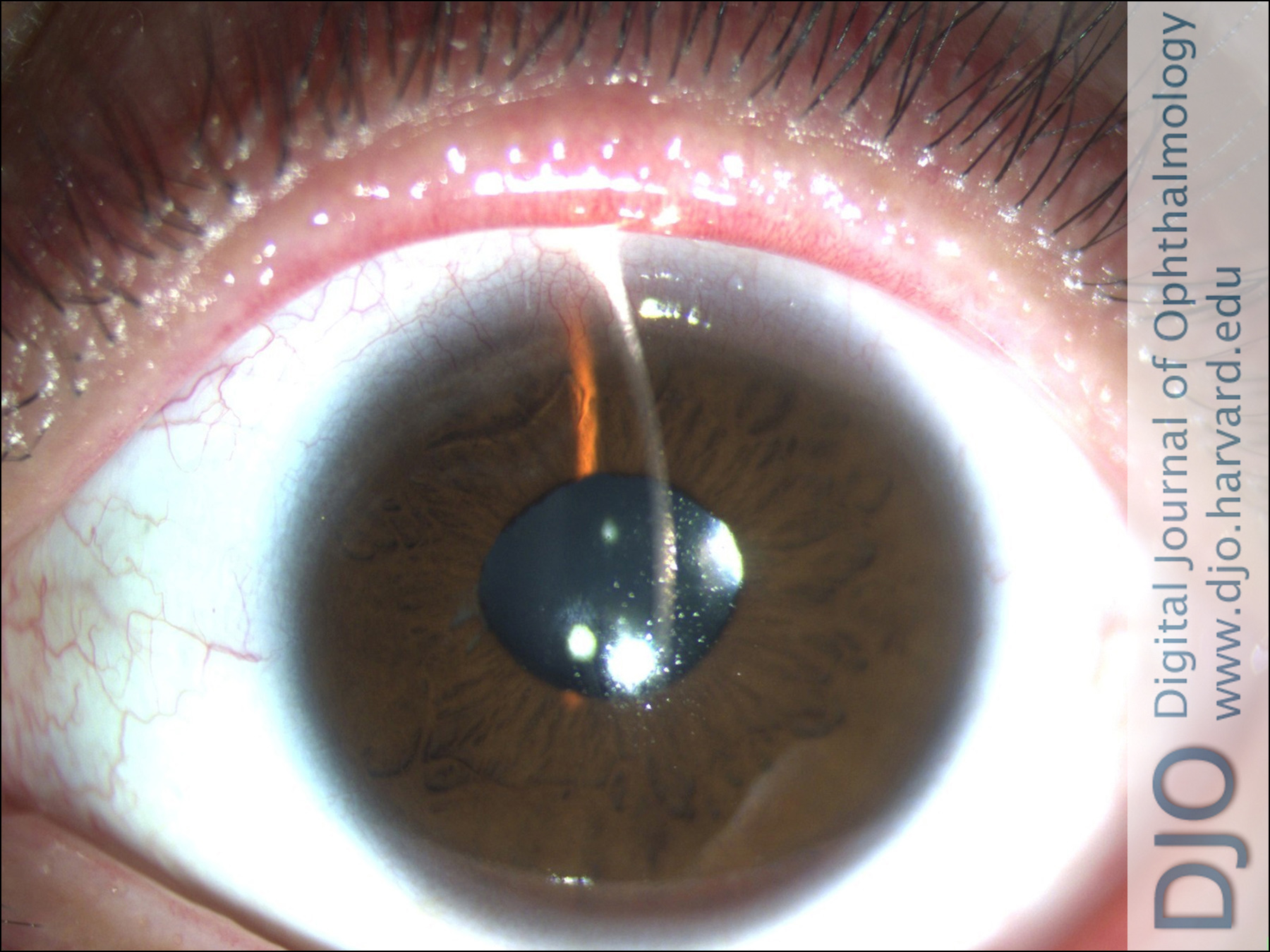
Ten months after ICL implantation, the patient returned to our clinic because of asymptomatic pupil deformation in his left eye (Figure 1). Distance visual acuity (corrected) in this eye was 16/20 with a refraction of +0.75 +0.75 ×170. Intraocular pressure (IOP) was 17 mm Hg. Slit-lamp microscopy of the left eye showed a clear cornea and partial dislocation of the ICL into the anterior chamber, with pupillary capture. No iris atrophy was observed. The patient denied history of trauma or strenuous activity. The ICL was successfully repositioned. On the first day after surgery, distance visual acuity (corrected) was 12/20, and IOP was 15 mm Hg. Slit-lamp microscopy revealed a clear cornea, a slightly, oval pupil and clear ICL in position (Figure 2). Vaulting was 310 μm on anterior segment optical coherence tomography. One month later, (corrected) distance visual acuity was 16/20, IOP was 15 mm Hg, and vaulting was 442 μm (Figures 3 and 4). | |
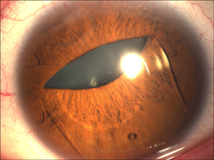
Figure 1
Anterior segment photograph showing partial dislocation of the implantable collamer lens (ICL) into the anterior chamber with pupillary capture.
|
|
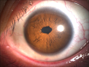
Figure 2
Anterior segment photographs of the left eye on postoperative day 1 showing a slightly oval pupil after ICL repositioning.
|
|
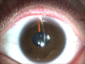
Figure 3
Anterior segment photographs of the left eye 1 month after surgery showing the well-positioned ICL with normal pupil.
|
|
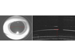
Figure 4
Anterior segment optical coherence tomograph of the left eye (Cirrus HD-OCT, Carl Zeiss Meditec AG, Jena, Germany) 1 month after surgery showing the normal vault.
|
|
| Discussion | Only 6 cases of postoperative ICL dislocation have been reported in the literature. In 4 cases, dislocation occurred after blunt trauma to the eye;(4-7) in 1 case, after occipital trauma.(8) The only reported case of spontaneous ICL dislocation was described in 2005 and was related to the rupture of the zonules in the inferior quadrants, with no displacement in the anterior chamber.(9)
To our knowledge, this is the first case of spontaneous dislocation of an ICL into the anterior chamber. Since neither direct nor indirect trauma was reported by the patient, the mechanism of dislocation had to involve a maximum pupillary dilation. We believe that drug-induced mydriasis may have played a role in this instance of ICL dislocation.
Olanzapine belongs to the class of benzodiazepines and is a new type of antipsychotic drug, whose main mechanism of action is to block serotonin 5-hydroxytryptamine (5-HT)2a and deoxyadenosine (DA) receptors. Buspirone is a nonbenzodiazepine anxiolytic agent that has a high affinity for serotonin 5-HT1a-receptor. Both drugs show an anticholinergic activity,(10-12) which can induce pupil dilation and cycloplegia. We speculate that both pupil dilation and cycloplegia may trigger ICL dislocation into the anterior chamber in combination with other factors, such as face-down head lowering and dark-induced mydriasis. This case suggests that systemic drugs with anticholinergic activity may represent a relative contraindication to ICL implantation.
Literature Search
PubMed was searched on December 7, 2019, for English-language results, using the following terms and combinations: pupillary capture AND ICL. | | | References | 1. Sanders DR, Vukich JA, Doney K, et al. U.S. Food and Drug Administration clinical trial of the implantable contact lens for moderate to high myopia. Ophthalmology 2003;110:255-66.
2. Packer M. The implantable collamer lens with a central port: review of the literature. Clin Ophthalmol 2018;12:2427-38.
3. Alfonso JF, Lisa C, Fernandez-Vega L, Almanzar D, Perez-Vives C, Montes-Mico R. Prevalence of cataract after collagen copolymer phakic intraocular lens implantation for myopia, hyperopia, and astigmatism. J Cataract Refract Surg 2015;41:800-805.
4. Morshifar M, Stagg BC, Muthappan V, Vasavada SA. Traumatic dislocation of implanted collamer phakic lens: a case report and review of the literature. Open J Ophthalmol 2014 May 30;8:824-6.
5. Song JS, Moon HS. Pupillary capture of implantable contact lens after blunt trauma. J Cataract Refract Surg 2005;31:1831-3.
6. Schmitz JW, McEwan GC, Hofmeister EM. Delayed presentation of traumatic dislocation of a visian implantable collamer lens. J Refract Surg 2012;28:365-7.
7. Espinosa-Mattar Z, Gomez-Bastar A, Graue-Hernandez EO, et al. DSAEK for implantable collamer lens dislocation and corneal decompensation 6 years after implantation. Ophthalmic Surg Lasers Imaging 2012;43:e68-72.
8. Kong J, Qin XJ, Li XY, Zhang JS, Qu B. Implantable collamer lens dislocation. Ophthalmology 2010;117:399.e1.
9. Kaufer RA, Kaufer GJ. Late subluxation of an ICL. J Cataract Refract Surg 2005;31:1254-5.
10. Chew ML, Mulsant BH, Pollock BG, et al. A model of anticholinergic activity of atypical antipsychotic medications. Schizophr Res 2006;88:63-72.
11. Mulsant BH, Gharabawi GM, Bossie CA, et al. Correlates of anticholinergic activity in patients with dementia and psychosis treated with risperidone or olanzapine. J Clin Psychiatry 2004;65:1708-14.
12. Kolasa K, Fusi R, Garattini S, Consolo S, Ladinsky H. Neurochemical effects of buspirone, a novel psychotropic drug, on the central cholinergic system. J Pharm Pharmacol 1982;34:314-7. | |
|
|
|
|
|
|
 Welcome, please sign in