|
|
 |
|
|
|
|
|
Self-inflicted injection of tattoo ink in the anterior chamber: a failed attempt to change the color of the eyes
Digital Journal of Ophthalmology
2018
Volume 24, Number 2
June 30, 2018
DOI: 10.5693/djo.02.2018.01.002
|
Printer Friendly
|
|
|

José D. Paulo, MD | Universidad Pontificia Bolivariana, Medellin, Colombia; Hospital Pablo Tobón Uribe, Medellin, Colombia; Universidad de Antioquia, Medellin, Colombia Juan Carlos Mejía-Turizo, MD | Universidad Pontificia Bolivariana, Medellin, Colombia Diana Carolina Montoya-Carrasquilla, MD | Universidad Pontificia Bolivariana Luisa Maria Bustamante, MD | Universidad Pontificia Bolivariana, Medellin, Colombia
|
|
|
| Abstract | | We report the case of a 29-year-old man, a tattoo artist by profession, with a history of schizophrenia, who was admitted to our hospital after injecting tattoo pigments in the anterior chamber of both eyes using a dermograph. The patient was diagnosed with penetrating ocular injury with secondary glaucoma, endotheliitis, and uveitis. Anterior chamber washout was performed immediately. At 4 months’ follow-up the patient had a visual acuity of 20/25 in each eye and 360° peripheral anterior synechiae; there still were traces of tattoo pigment at the trabecular meshwork and endothelial corneal layer. | | | Introduction | | Conjunctival tattooing is an extreme form of body modification and part of an increasing trend in unusual tattoos.(1) The technique makes use of an ink-coated needle that oscillates about 100 times per second, depositing ink pigments 1.5–2 mm below the surface. Reports of tattoo-related adverse reactions and accidental injuries, mostly on the skin, are frequent. We report a case of self-inflicted injections of tattoo ink in the anterior chamber by an individual in a failed attempt to change the color of his eyes. | | | Case Report | A 29-year-old man, a tattoo artist by profession, with a history of schizophrenia, was admitted to our hospital after injecting tattoo pigment into the anterior chamber of both of his eyes using a dermograph. The patient presented with pain and severe decrease in visual acuity. On examination, visual acuity was hand motions in the right eye and counting fingers in the left eye. Both eyelids displayed mild edema, conjunctival chemosis, and ciliary injection. There was severe corneal edema with multiple self-sealing punctate wounds in the inferior area of the cornea. The anterior chamber was filled with green ink (see Figure 1). Intraocular pressure (IOP) was 35 mm Hg in the right eye and 44 mm Hg in the left eye. Due to the dense hue of the greenish substance, it was not possible to observe more detail. The patient was diagnosed with penetrating ocular injury with secondary glaucoma, endothelitis, and uveitis. Anterior chamber washout was performed immediately. Topical treatment with ciprofloxacin, prednisolone, and tropicamide was initiated, and a single intravenous dexamethasone dose was administered. The patient was discharged from the hospital the next day, with an IOP of 32 mm Hg in the right eye and 36 mm Hg in the left eye; topical briminidine and timolol twice daily was prescribed. Psychiatric evaluation prior to discharge revealed no cause to detain the patient and no suicidal ideation.
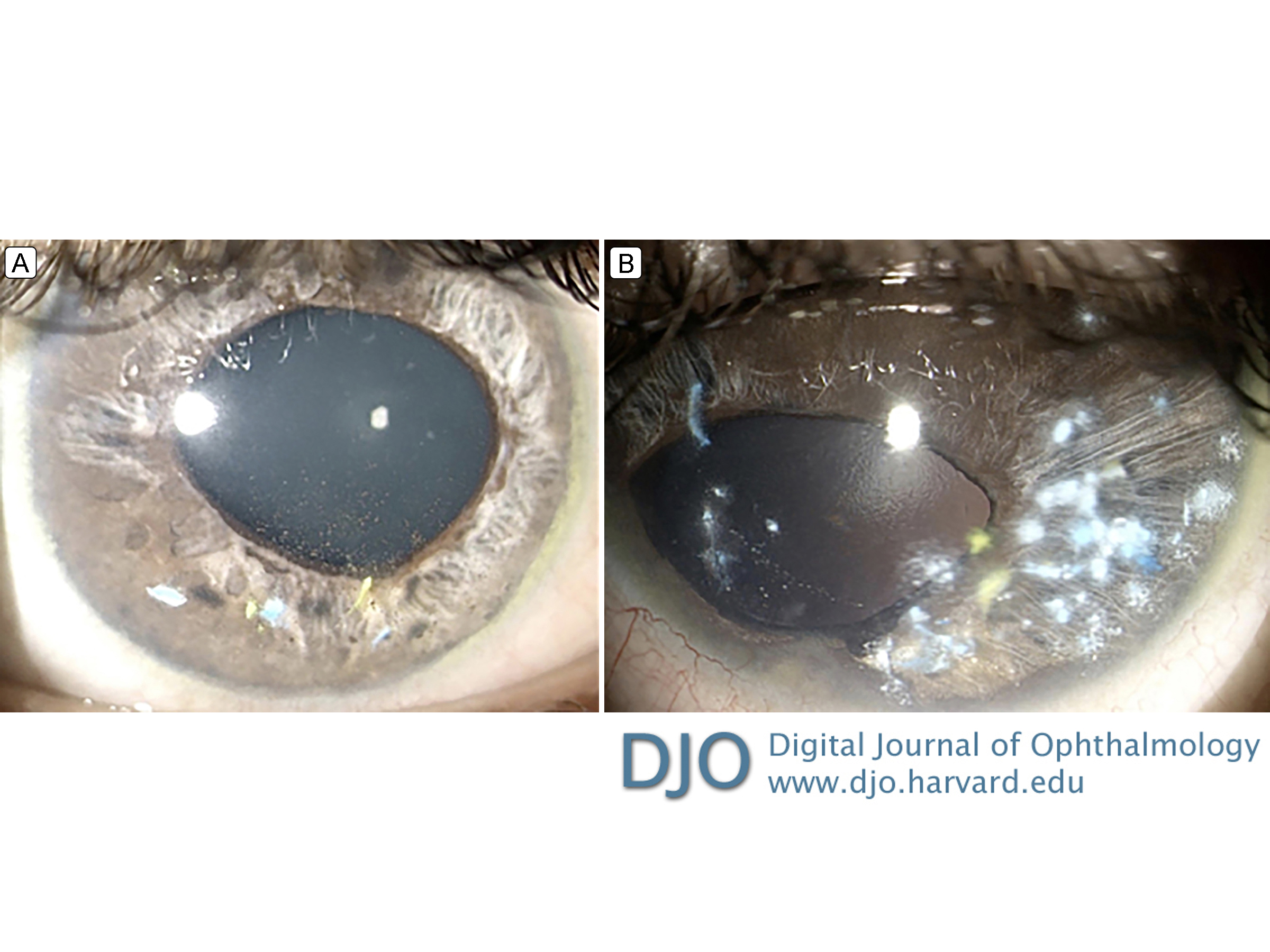
At 4 months’ follow-up, the patient had a visual acuity of 20/25 in each eye, leucomas in the inferotemporal zone of both corneas, 360° peripheral anterior synechiae, and iris atrophy. There still were traces of tattoo pigment at the trabecular meshwork and endothelial corneal layer (Figure 2). IOP measured 12 mm Hg in each eye, with topical antiglaucoma medications. There were normal-appearing optic nerve heads on fundus examination, without increase of the excavation or alterations of the neuroretinal rim. The maculae, vessels, and periphery of both eyes were normal. Endothelial cell counts were performed and were decreased for his age (right eye, 2086 cells/mm2; left eye, 1869 cells/mm2 (normal for age, 2520 cells/mm2).
| |

Figure 1
Biomicroscopy of right eye (A) and left eye (B) at admission showing generalized conjunctival chemosis, corneas with edema and multiple inferior perforations, and green tattoo ink filling the anterior chamber.
|
|

Figure 2
Biomicroscopy of the right eye (A) and left eye (B) at 4 months’ follow-up. Tattoo ink is seen in the endothelium and anterior chamber angle along with generalized atrophy of the iris and anterior peripheral synechiae, worse in the left eye.
|
|
| Discussion | Few cases describing the effects of tattoo ink in the eye have been reported,(2-4) and we found only 2 intraocular penetrations with severe complications (including inflammation, retinal toxicity, glaucoma, and retinal detachment) produced by chalcosis.(5,1) Tattoo artists use azo- and polycyclic pigments containing copper, chromium, and cobalt to create all colors of the visible spectrum. Copper, in particular, can cause of severe ocular inflammation.(1) Toxic anterior segment syndrome (TASS) is usually a complication of anterior segment eye surgery or accidental penetration of the eyeball;(6) the severe inflammatory reactions of our patient were compatible with TASS. The clinical picture was characterized by the TASS-like features of total corneal edema due to severe endothelitis, fibrinous reaction in the anterior chamber, and ocular hypertension due to damage to the trabecular meshwork, associated with minimal pain, and without vitreous involvement. Management with anterior chamber irrigation to eliminate most of the pigment, topical and systemic steroids, and glaucoma medication proved adequate to control the toxicity and achieve good visual acuity in the early follow-up period, but damage to the trabecular meshwork and corneal endothelium necessitated long-term management. Moreover, without electroretinography and serial follow-up, we cannot be certain whether this patient had any retinal toxicity from possible calcosis in addition to TASS.
Literature Search
PubMed was searched in June 2017 using the following terms: toxic anterior segment syndrome; coloring agents AND adverse effects; tattooing AND methods, adverse effects; corneal opacity AND therapy; eye foreign bodies; ophthalmic solutions AND adverse effects.
| | | References | 1. Jalil A, Ivanova T, Bonshek R, Patton N. Unique case of eyeball tattooing leading to ocular penetration and intraocular tattoo pigment deposition. Clin Experiment Ophthalmol. 2015;43:594-6.
2. Panda A, Mohan M, Chawdhary S. Corneal tattooing—experiences with “lamellar pocket procedure.” Indian J Ophthalmol 1984;32:408-11.
3. Pradhan S, Das M, Panigrahi AK, Prajna NV. Severe conjunctival reaction following attempted corneal tattooing. JAMA Ophthalmol 2015;133:854.
4. Van der Velden/Samderubun EM, Kok JH. Dermatography as a modern treatment for coloring leucoma corneae. Cornea 1994;13:349-53.
5. Cruz NF, Santos KS, Farah ML, Felberg S. Conjunctival tattoo with inadvertent globe penetration and associated complications. Cornea 2017;36:625-7.
6. Mamalis N, Edelhauser HF, Dawson DG, Chew J, LeBoyer RM, Werner L. Toxic anterior segment syndrome. J Cataract Refract Surg 2006;32:324-33.
| |
|
|
|
|
|
|
 Welcome, please sign in