|
|
 |
|
|
|
|
|
Stalagmite-like preretinal inflammatory deposits in vitrectomized eyes with posterior uveitis
Digital Journal of Ophthalmology
2017
Volume 23, Number 1
February 18, 2017
DOI: 10.5693/djo.02.2016.02.002
|
Printer Friendly
Download PDF |
|
|

Yoshihiro Yonekawa, MD | Massachusetts Eye and Ear Infirmary, Boston, Massachusetts; Boston Childrens Hospital, Boston, Massachusetts Ashkan M. Abbey, MD | Texas Retina Associates, Dallas Lily van Laere, MD | Department of Ophthalmology, William Beaumont Hospital, Royal Oak, Michigan Ankoor R. Shah, MD | Retina Consults of Houston, Houston, Texas Benjamin J. Thomas, MD | Florida Retina Institute, Jacksonville Alan J. Ruby, MD | Department of Ophthalmology, William Beaumont Hospital, Royal Oak, Michigan Associated Retinal Consultants, Royal Oak, Michigan Lisa J. Faia, MD | Department of Ophthalmology, William Beaumont Hospital, Royal Oak, Michigan Associated Retinal Consultants, Royal Oak, Michigan
|
|
|
| Abstract | | We report a new clinical sign of vitreous inflammation in patients with posterior uveitis: spectral-domain optical coherence tomography identified stalagmite-like, discrete, diffusely distributed, hyperreflective, preretinal deposits in previously vitrectomized eyes of 2 patients during flares of posterior uveitis. The extent of the deposits correlated with disease activity. The underlying primary diseases encountered were necrotizing retinochoroiditis secondary to toxoplasmosis and primary central nervous system lymphoma. | | | Introduction | | Advanced imaging modalities have enhanced our management of posterior uveitides. From optical coherence tomography (OCT) for examining macular edema to fundus autofluorescence for gauging disease activity in white dot syndromes, developments in diagnostic imaging have been clinically meaningful and have improved our understanding of these complex ophthalmic disorders. We describe a new spectral domain OCT (SD-OCT) sign of vitreous inflammation observed in 2 patients with vitrectomized eyes and flares of posterior uveitis who were seen at Associated Retinal Consultants, William Beaumont Hospital. The stalagmite-like lesions are multifocal and discrete preretinal hyperreflective deposits that resemble the elevated speleothems. | | | Case Report | Case 1
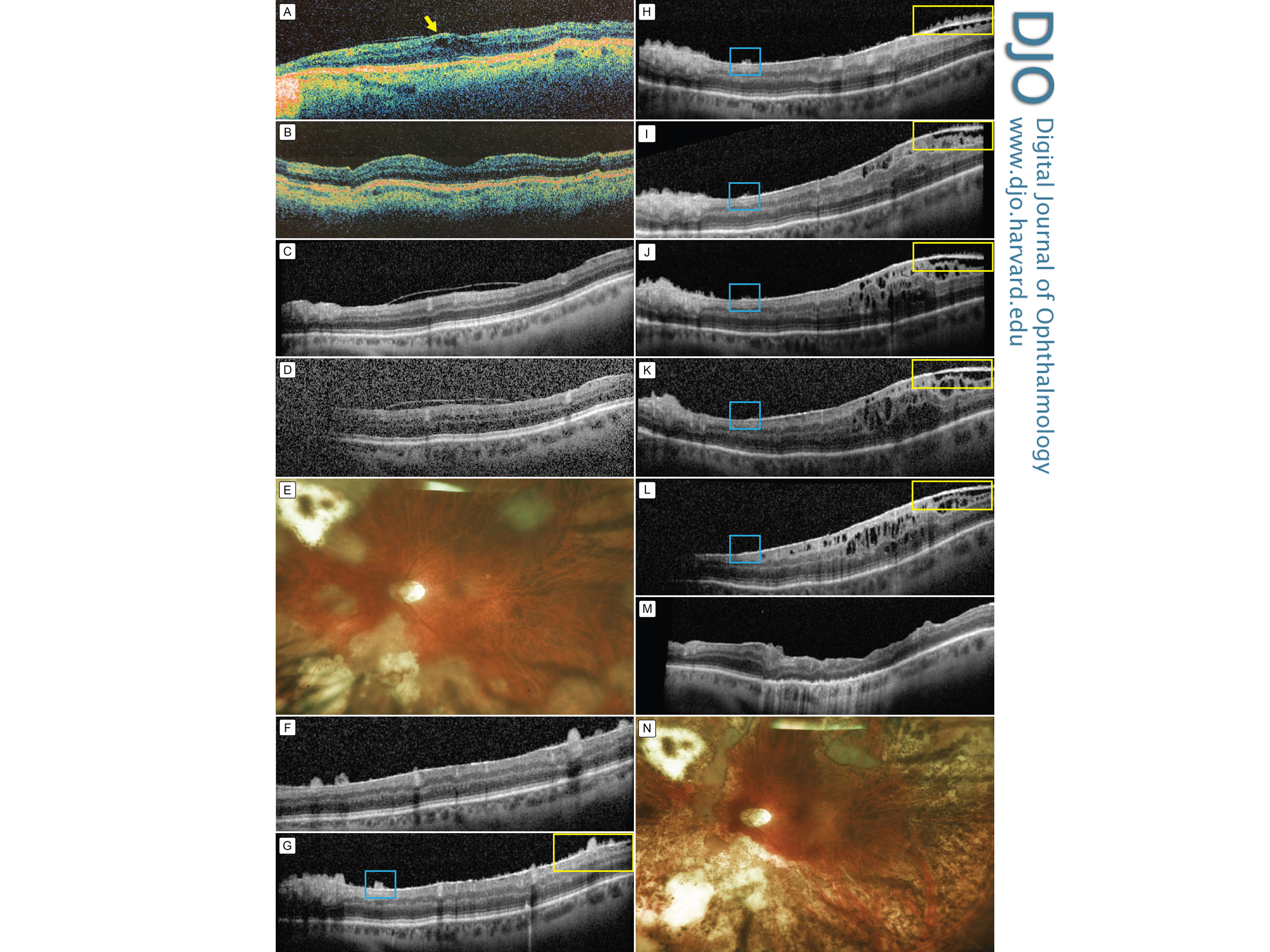
A 69-year-old man with a history of idiopathic panuveitis in both eyes was referred for further management. On examination, visual acuity was 20/30 in the right eye and 20/40 in the left eye. Intraocular pressure (IOP) was 15 mm Hg in the right eye and 11 mm Hg in the left eye. The anterior segments of both eyes were quiet. Fundus examination showed changes in the macular retinal pigment epithelium (RPE) and scattered inferior peripheral chorioretinal atrophy without pigmentation in both eyes. Time-domain OCT (TD-OCT; Stratus OCT; Carl Zeiss Meditec, Dublin, CA) showed normal retinal anatomy in the right eye but mild intraretinal fluid in the left eye (Figure 1A). An inflammatory and infectious workup was unrevealing. The edema resolved with topical steroid treatment (Figure 1B). The first SD-OCT (Spectralis SD-OCT; Heidelberg Engineering, Heidelberg, Germany), obtained 1 year after presentation, was unremarkable (Figure 1C). Over the next several months, the patient accumulated vitreous debris through syneresis that became visually significant, with visual acuity declining to 20/200. Shadowing by the debris was seen on SD-OCT, but the retinal architecture remained unaffected (Figure 1D). A diagnostic pars plana vitrectomy and membrane peeling were performed, but the vitreous analyses were unrevealing.
Visual acuity improved to 20/60 at the 1-week postoperative visit but declined to 20/100 at 1 month postoperatively, and the IOP was found to be elevated at 28 mm Hg. Fundus examination revealed a multifocal necrotic retinitis with peripheral vitritis (Figure 1E). SD-OCT showed diffusely scattered, discrete, hyperreflective stalagmite-like vitreous condensations that had deposited on the retinal surface (Figure 1F); these lesions were not appreciated on clinical examination or fluorescein angiography. A suspicion for a viral retinitis prompted an anterior chamber tap and intravitreal injections of ganciclovir and foscarnet. The patient was also started on oral prednisone and valacyclovir.
Polymerase chain reaction (PCR) amplification was negative for varicella zoster virus, herpes simplex viruses 1 and 2, and cytomegalovirus; however, subsequent analyses revealed PCR positivity for Toxoplasma gondii. The diagnosis was reformulated as toxoplasmosis retinochoroiditis presenting as a subacute retinal necrosis, and the patient was treated with pyrimethamine, sulfadiazine, prednisone, and folic acid. The retinitis, vitritis, and preretinal deposits gradually improved, and the patient was maintained on sulfamethoxazole-trimethoprim (Figures 1G-L). The patient subsequently underwent removal of a recurrent epiretinal membrane (Figure 1M). The preretinal deposits, retinitis, and vitritis have not recurred during the 13 months of follow-up (Figure 1N).
Case 2
A 71-year-old woman with a history of primary central nervous system (CNS) B-cell lymphoma, treated 1 year before presentation with intravenous methotrexate and rituximab, presented with floaters in the right eye. Visual acuity was 20/30 in each eye, and IOP was normal. Examination revealed 2+ vitreous cells, trace RPE changes, and no other retinal or choroidal lesions. TD-OCT revealed shadowing from the vitreous debris but no other abnormalities (Figure 2A). A diagnostic vitrectomy revealed a single atypical large lymphocyte on cell cytology and a monoclonal kappa B cell population. No CNS lesions were seen on MRI. The patient declined further therapies. Visual acuity improved to 20/30, and the eye entered a period of remission. TD-OCT at postoperative month 1 showed normal retinal architecture (Figure 2B).
On follow-up examination 1.5 years later, the patient reported a gradual recurrence of floaters in the right eye. Visual acuity was stable at 20/40 but new vitreous cells were noted on examination. SD-OCT revealed heterogeneously sized stalagmite-like hyperreflective preretinal deposits (Figure 2C). Serial brain MRIs remained clear, without evidence of recurrence. The patient was closely observed, and though the preretinal deposits fluctuated, they gradually improved over several months without intervention (Figures 2D-H). Visual acuity after 23 months decreased to 20/60 and associated degenerative changes were seen in the outer retina (Figures 2D-H). At 23-months’ follow-up, the OCT remained unremarkable, but the brain MRI showed recurrence of the intracranial lymphoma, and the patient was started on systemic treatment. Her ocular examination remained quiescent, with no signs of intraocular inflammation. | |

Figure 1
Stalagmite-like preretinal deposits in a vitrectomized eye with toxoplasmosis retinochoroiditis. A, Time domain optical coherence tomography (TD-OCT) demonstrating mild macular edema (arrow) at time of initial referral. B, TD-OCT 5 months later showing good response to topical steroid therapy. C, The first spectral domain OCT (SD-OCT) was obtained 5 months subsequently and shows a partially attached posterior hyaloid and temporal epiretinal membrane. The selected SD-OCT B-scan is one disc diameter superior to the fovea, illustrating the subsequent dynamic preretinal deposits best. D, Over the course of 3-4 months, the patient gradually developed worsening floaters; SD-OCT showed optically significant vitreous debris. E, One month after a pars plana vitrectomy with an unrevealing vitreous biopsy, the patient developed a multifocal necrotic retinitis with vitritis, as seen in this widefield photograph. F, SD-OCT showed diffuse stalagmite-like hyperreflective preretinal deposits. G, Three weeks after intravitreal antivitral treatment and oral steroids the preretinal deposits were slightly smaller and with sharper edges. Subsequent B-scans are of the identical cut, and the colored boxes follow the progression of individual lesions. H, 1 month later, the preretinal deposits are significantly smaller after initiating treatment for toxoplasmosis. I, Another month later, the deposits are almost revolved, but there is increasing thickening of the epiretinal membrane (ERM) and associated tractional changes. J-L, The improvement in the deposits and concurrent worsening of the ERM continue 1 (J), 3 (K), and 6 (L) months subsequently. M, Edema resolved after pars plana vitrectomy with membrane peeling. N, Retinitis had not recurred during 13 months of follow-up, leaving patches of quiescent chorioretinal atrophy.
|
|
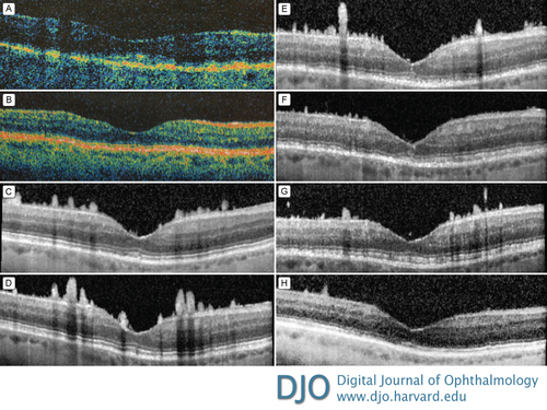
Figure 2
Stalagmite-like preretinal deposits in a vitrectomized eye with intraocular lymphoma. A, TD-OCT at initial presentation showing shadowing from vitreous debris. B, TD-OCT showing normal retinal architecture 1 month after a diagnostic vitrectomy. C, SD-OCT 1.5 years later shows diffuse, multiple, elongated, hyperreflective preretinal deposits. D-H, Deposits gradually improve without treatment over the course of the subsequent 8 (D), 10 (E), 12 (F), 16 (G), and 19 (H) months.
|
|
| Discussion | SD-OCT has emerged as a valuable and versatile diagnostic tool in the management of all anatomic layers of the posterior segment in patients with uveitis, including grading of vitreous haze,(1) vitreomacular interface abnormalities such as epiretinal membranes and vitreomacular traction,(2) and deeper abnormalities within and below the neurosensory retina, such as cystoid macular edema and choroidal neovascularization. The present study describes a new vitreoretinal interface SD-OCT finding that appears to correlate with inflammatory activity. The lesions are hyperreflective discrete preretinal deposits distributed throughout the macula on the retinal surface. The shapes and sizes are heterogeneous: some are large—up to approximately 200 µm—whereas others are barely visible. Some are lobules of hyperreflective material, whereas others are needle shaped. Overall, most have broader bases with a distinct tapering towards the vitreous cavity, which resemble stalagmite formations.
Both patients had underlying posterior uveitis at baseline: presumed idiopathic in case 1 and intraocular lymphoma in case 2. In both eyes, the preretinal deposits occurred during inflammatory activity after the eyes had undergone vitrectomy. We hypothesize that these deposits are aggregations of inflammatory cells that adhere to the retinal surface. The lesions did not have a noticeable predilection for the inferior macula, so the cellular aggregates were adhering to the retinal surface without substantial gravity dependence. This is unlike the common presentation of vitreous opacities, where the cells normally circulate in the midvitreous or settle in the inferior vitreous base.
There was subtle phenotypic variability between the 2 cases. In case 1, the patient developed a necrotizing retinochoroiditis from toxoplasmosis. The subacute intraocular inflammation initially caused larger aggregates of preretinal deposits (Figure 1E), which subsequently became smaller and sharper (Figures 1F-I) until almost completely resolved (Figure 1K). A more flattened preretinal oval lesion that appears to follow a similar pattern of resolution with treatment has been noted, although previous authors do not specify the vitrectomy status of these patients, which may account for the difference in shape.(3) The linear improvement in our case similar to other cases was likely a result of the treatment of the toxoplasmosis and associated inflammation.
In case 2, the preretinal deposits were dynamic. Both the height and width of the stalagmite-like lesions increased between the initial (Figure 2C) and second (Figure 2D) SD-OCT then gradually improved, but with some degree of fluctuation. This patient’s diagnosis was vitreous inflammation in an eye with a history of intraocular lymphoma that had been in remission after a diagnostic vitrectomy without treatment. There were no other signs of active lymphoma, but reactivation could not be ruled out, because further intervention was declined; therefore, the possibility that the preretinal deposits consisted of lymphoma cells rather than pure aggregations of leukocytes cannot be ruled out. Interestingly, there was a discordance between her intraocular findings and her systemic disease: there was no intraocular involvement during the initial CNS lymphoma; the preretinal deposits presented 1.5 years later during a period of remission; 2 years after the appearance of the intraocular inflammation that spontaneously resolved, the CNS lymphoma recurred, but there was no intraocular activity.
These stalagmite-like preretinal deposits have not been previously described. The most similar entity was reported by Paulus et al,(4) who recently described 2 patients with isolated prefoveal vitreous condensations. One patient was a child with intermediate uveitis in the setting of a positive purified protein derivative skin test, who developed a single spirelike preretinal deposit seen on SD-OCT during a recurrence. The second case was a woman previously treated for primary CNS lymphoma, who presented with a single prefoveal spirelike deposit. Another report described five cases of ocular toxoplasmosis that demonstrated preretinal ovoid lesions discrete from a chorioretinal toxoplasmosis scar that appeared to develop and resolve with treatment.(5)
The vitreous can assume many formations in eyes with chronic inflammation. Classic vitritis is the most common presentation, but other forms of inflammatory vitreous debris include snowballs, snowbanking, vitreous cylinders, the recent single spirelike prefoveal deposits,(4) and with the current report, stalagmite-like diffuse preretinal deposits. Snowballs and snowbanking occur most frequently in intermediate uveitis; vitreous cylinders are relatively nonspecific columns of inflamed collagen fibrils;(5) and spirelike prefoveal deposits appear to occur in nonvitrectomized eyes with posterior uveitis.(4) In the present report of diffuse stalagmites, the unifying clinical history is inflammation in previously vitrectomized eyes. The inflammatory cells are able to move freely in a vitrectomized eye via Brownian-like motion, and therefore likely assume a diffusely distributed pattern. The uniformity of the distribution was appreciated on the SD-OCT scans, where the stalagmite lesions were similar in number in the superior and inferior scans. This is in contrast to less freely mobile inflammatory vitreous deposits in nonvitrectomized eyes, where vitreous debris tends to accumulate inferiorly or at the fovea.(4) The specific affinity of the inflammatory cells to the retinal surface may occur for a number of reasons, including the upregulation of cellular adhesion molecule expression seen in experimental autoimmune uveoretinitis.(6) Beyond retinal surface adhesion, given the multicellular size of these stalagmite-like lesions, leukocyte aggregation is likely involved. The exact mechanism for the propensity for leukocytes to aggregate together during inflammation is unknown, but some have proposed humoral factors that are upregulated during inflammation, such as fibrinogen, to play a role in the induction and/or maintenance of the increased adhesive properties.(7) However, there are currently no studies examining leukocyte-leukocyte adhesion in ocular inflammation and, surprisingly, no strong evidence in the overall medical literature. Furthermore, we are making an assumption that the stalagmites are inflammatory aggregates, but this remains an imaging finding without a clear mechanism until histologic studies are performed in the future.
Literature Search
A PubMed search was conducted on June 4, 2015, for English-language results using the following search terms: optical coherence tomography and uveitis. | | | References | 1. Keane PA, Karampelas M, Sim DA, et al. Objective measurement of vitreous inflammation using optical coherence tomography. Ophthalmology 2014;121:1706-14.
2. Nicholson BP, Zhou M, Rostamizadeh M, et al. Epidemiology of epiretinal membrane in a large cohort of patients with uveitis. Ophthalmology 2014;121:2393-8.
3. Goldenberg D, Goldstein M, Loewenstein A, Habot-Wilner Z. Vitreal, retinal, and choroidal findings in active and scarred toxoplasmosis lesions: a prospective study by spectral-domain optical coherence tomography. Graefes Arch Clin Exp Ophthalmol 2013;251:2037-45.
4. Paulus YM, Wong IG, Sanislo S, Moshfeghi DM. Prefoveal vitreous condensation in chronic inflammation. Ophthalmic Surg Lasers Imaging Retina 2014;45:447-50.
5. Roizenblatt J, Grant S, Foos RY. Vitreous cylinders. Arch Ophthalmol 1980;98:734-9.
6. Xu H, Forrester JV, Liversidge J, Crane IJ. Leukocyte trafficking in experimental autoimmune uveitis: breakdown of blood-retinal barrier and upregulation of cellular adhesion molecules. Invest Ophthalmol Vis Sci 2003;44:226-34.
7. Urbach J, Lebenthal Y, Levy S, et al. Leukocyte adhesiveness/aggregation test (LAAT) to discriminate between viral and bacterial infection in children. Acta Paediatr 2000;89:519-22. | |
|
|
|
|
|
|
 Welcome, please sign in