|
|
 |
 |
 |
 |
|
|
Retinal metastasis from unknown primary: diagnosis, management, and clinicopathologic correlation
Digital Journal of Ophthalmology
2015
Volume 21, Number 4
October 5, 2015
DOI: 10.5693/djo.02.2015.04.004
|
Printer Friendly
Download PDF |
|
|


Kenneth J. Taubenslag, MPhil | Vanderbilt Eye Institute, Vanderbilt University Medical Center, Nashville, Tennessee Stephen J. Kim, MD | Vanderbilt Eye Institute, Vanderbilt University Medical Center, Nashville, Tennessee Albert Attia, MD | Department of Radiation Oncology, Vanderbilt University Medical Center; Vanderbilt-Ingram Cancer Center, Vanderbilt University Medical Center Ty W. Abel, MD, PhD | Vanderbilt-Ingram Cancer Center, Vanderbilt University Medical Center; Department of Pathology, Microbiology, and Immunology, Vanderbilt University Medical Center Hilary Highfield Nickols, MD, PhD | Vanderbilt-Ingram Cancer Center, Vanderbilt University Medical Center; Department of Pathology, Microbiology, and Immunology, Vanderbilt University Medical Center Kristin K. Ancell, MD | Vanderbilt-Ingram Cancer Center, Vanderbilt University Medical Center; Medical Oncology, Department of Medicine, Vanderbilt University Medical Center Anthony B. Daniels, MD, MSc | Vanderbilt Eye Institute, Vanderbilt University Medical Center, Nashville, Tennessee; Department of Radiation Oncology, Vanderbilt University Medical Center; Vanderbilt-Ingram Cancer Center, Vanderbilt University Medical Center; Department of Cancer Biology, Vanderbilt University, Nashville, Tennessee
|
|
|
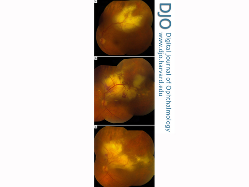
| Abstract | | A 75-year-old man was incidentally found to have a yellow-white retinal lesion with scattered hemorrhages. He was empirically treated elsewhere for viral retinitis without resolution and later transferred to the Vanderbilt Eye Institute, where retinal biopsy with silicone oil tamponade showed retinal metastasis. He had no prior history of cancer, and multiple systemic imaging evaluations failed to identify a primary site. Histopathology and immunohistochemistry of the biopsy were consistent with non-small-cell lung carcinoma. Due to the radiation-attenuating properties of silicone oil, the patient underwent silicone oil removal prior to receiving external beam radiotherapy (EBRT). The retinal metastasis responded completely to EBRT, and at final follow-up, 18 months after initial presentation, no primary tumor has been identified. | | | Introduction | | Although uveal metastases are relatively common, metastasis to the retina is very rare, with only 29 cases of carcinoma metastatic to the retina reported in the literature.(1-4) Of these, no primary site could be identified in 3 cases.(1-2) Many other entities, including vascular occlusions, infectious retinitis, and lymphoproliferative disorders can masquerade as retinal metastasis, making this entity a diagnostic challenge, especially in patients with no prior history of cancer or in patients in whom no primary tumor can be found. We present a rare case of retinal metastasis in which no primary tumor could be identified. Initial misdiagnosis prior to referral led to delay in correct treatment, progression of the lesion, and visual loss. Retinal biopsy with HistoGel (Thermo Fisher Scientific, Waltham, MA) tissue-processing and immunohistochemical analysis ultimately led to the correct diagnosis.(5) We discuss the approach to retinal metastasis from unknown primary, the role of biopsy, and the challenges and implications of radiation-attenuating vitreous substitutes such as silicone oil. | | | Case Report | A 75-year-old male smoker was incidentally noted on routine examination elsewhere to have a poorly demarcated yellow-white retinal lesion superotemporally in the left eye, with intraretinal hemorrhages and increased vascular tortuosity along the superotemporal arcade (Figure 1A). Visual acuity was 20/25 in this eye and 20/100 in the contralateral eye from preexisting corneal scar. Neoplasm was entertained as a possible diagnosis. A confirmatory biopsy was recommended, but the patient declined. Metastatic workup, including positron emission tomography computed tomography (PET-CT) was negative for primary tumor elsewhere. The patient was diagnosed with branch retinal vein occlusion (BRVO) with Coats-like reaction.
Five months later, he noticed blurred vision in the left eye, and examination showed 1+ vitreous cells. Serum serologies were negative for Toxoplasma gondii and active varicella zoster. Herpes simplex virus titers were mildly elevated, and a presumptive diagnosis of infectious retinitis was made. Acyclovir was started by the treating ophthalmologist, but visual acuity in the left eye continued to decrease, from 20/25 to 20/60 over the next month.
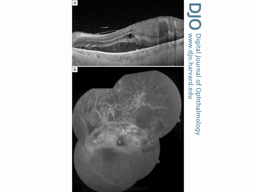
The patient was referred to the Retina Service at the Vanderbilt Eye Institute for further evaluation and management. The superotemporal yellowish retinal lesion was noted to have expanded. Optical coherence tomography (OCT) revealed thickening and disorganization of the retinal layers, with no apparent involvement of the subretinal space or underlying choroid (Figure 2A). Because of concern for secondary retinal malignancy, the patient underwent 23-gauge pars plana vitrectomy and retinal biopsy with endolaser retinopexy around the biopsy site and silicone oil tamponade. Histopathologic examination revealed clusters of epithelioid cells with eosinophilic to amphophilic cytoplasm, pleomorphic nuclei, prominent nucleoli, and numerous mitoses. Immunohistochemical evaluation was positive for cytokeratins AE1/3 and for thyroid transcription factor-1 (TTF-1); immunostains were negative for S100, Melan-A, CD20, and CD45, consistent with metastatic carcinoma of lung or thyroid origin (Figure 3). HistoGel tissue-processing medium utilized in the laboratory of the Division of Neuropathology allowed for successful examination of this very small biopsy specimen.
The patient was referred to the Ocular Oncology Service at the Vanderbilt Eye Institute. The retinal lesion had continued to expand, with abutment of the optic nerve head and presumed nerve invasion, with concomitant decrease in vision to counting fingers (Figure 1B). Repeat systemic imaging with CT chest, abdomen and pelvis with contrast, MRI of the brain and orbits and whole body PET scan failed to demonstrate a primary source, one year after the initial negative systemic imaging evaluation. Complete blood count remained within normal limits. A diagnosis of non-small cell lung carcinoma was made based on the histopathology, immunohistochemistry, and the absence of a thyroid lesion. Three weeks after the initial retinal biopsy, and after completion of the systemic evaluation, the patient underwent silicone oil removal, repeat endolaser, and 20% SF6 gas tamponade. Ten days later (Figure 1C), the bubble had decreased sufficiently to allow for tumor measurements by B-scan ultrasonography, which revealed a height of 3.61 mm, confined to the retina. He underwent three-dimensional conformal radiotherapy to the left globe (30 Gy in 10 fractions).
On examination 1 month after radiotherapy, the tumor had completely regressed and was no longer visible within the retina or adjacent to the nerve. However, the retina developed multiple breaks within the area of prior tumefaction, and rhegmatogenous retinal detachment developed. Visual acuity remained poor. Despite the rapid growth and progression of the retinal metastasis prior to treatment, no primary tumor site has become evident on systemic imaging more than 18 months after the retinal lesion was initially detected. | |

Figure 1
A, Fundus photography at time of presentation to Vanderbilt Eye Institute, 9 months after initial presentation, showing superotemporal yellow-white retinal lesion, hemorrhages, and vascular dilation and tortuosity. B, After retinal biopsy and silicone oil tamponade, there was continued growth of the lesion, now with juxtapapillary involvement, and visual acuity has dropped to hand motions. C, After removal of silicone oil, the lesion continued to grow and visual acuity remained hand motions.
|
|

Figure 2
A, Optical coherence tomography shows retinal thickening and disorganization of the retinal layers, with no apparent involvement of the subretinal space or underlying choroid. B, Fluorescein angiogram showing retinal hyperfluorescent lesion and vascular dilation with vessel staining.
|
|

Figure 3
Histopathology of retinal biopsy tissue. A, High-power magnification (600×) showing clusters of epithelioid cells with large, pleomorphic nuclei and prominent nucleoli. B-C, Immunohistochemistry shows tumor cell positivity for AE1/3 cytokeratin (B) in a membranous distribution and TTF-1 (C) in a nuclear distribution.
|
|
| Discussion | Cancer metastatic to the retina is a rare and challenging diagnosis, even more so in patients without a history of malignancy or in patients in whom a primary tumor source cannot be identified. In contrast to uveal metastases, which are quite common, only 29 cases of carcinoma metastatic to the retina have been reported. Of these, the primary site could be identified in 26 patients: 9 from gastrointestinal tract, 9 from lung cancer, 7 from breast adenocarcinoma, and 1 from the genitourinary tract.(1-4)
There are only 3 previous reported cases of retinal metastasis from unknown primary, and these present a diagnostic and therapeutic challenge.(1-2) Unlike choroidal metastases, retinal metastases rarely present with bilateral or multifocal lesions. Initial misdiagnosis leads to a delay for a mean interval of 5 months.(4) Common masquerading diagnoses include choroidal metastasis, primary choroidal melanoma, ocular lymphoma, focal retinitis, and vaso-occlusive disorders. Our patient carried several of these diagnoses (branch retinal vein occlusion, viral retinitis, vitreoretinal lymphoma) during the year between his initial presentation and our final correct diagnosis.
Ultimately, definitive diagnosis in our case was achieved with pathologic examination of a retinal biopsy specimen. Immunohistochemistry was positive for AE1/3 and TTF-1 suggesting lung or thyroid as the primary site. Coassin et al reported a similar immunohistochemical signature in a case of retinal metastasis of unknown primary.(2) Both adenocarcinoma and small cell carcinoma of the lung may express TTF-1, though squamous cell carcinoma typically does not. Extrapulmonary small cell carcinomas, but not adenocarcinomas, may also express TTF-1. Ultimately, the clear absence of thyroid tumor, and the histologic cytomorphology that was not consistent with a small cell tumor, allowed the diagnosis of metastatic, non-small cell lung adenocarcinoma. In addition, while 24 cases of thyroid cancer metastatic to the uvea have been reported,(6-8) retinal metastasis from thyroid primary has never been reported.
EBRT therapy was hindered by the presence of silicone oil in the vitreous cavity. Silicone oil was initially selected as a tamponade agent at the time of biopsy because vitreoretinal lymphoma was a consideration, and methotrexate can be injected into a silicone oil–containing eye, whereas dosing is difficult and diffusion properties altered by a vitrectomized or gas-containing cavity. In addition, the visual acuity in the contralateral eye was poor due the presence of a corneal scar, and silicone oil permitted the patient better vision than gas would have allowed.
A number of studies have examined the use of radiation-attenuating vitreous substitutes such as silicone oil for the prevention of radiation retinopathy after globe-conserving brachytherapy for uveal melanoma.(9-11) Replacing the radiation-attenuating oil with 20% SF6 ensured delivery of a predictable, unattenuated radiation dose to the tumor. Because the retina showed signs of fibrosis and traction, additional tamponade was desired after the supplemental endolaser. 20% SF6 was selected so that rapid aqueous replacement of the gas would allow for tumor measurements to be obtained quickly and EBRT planning and treatment performed with minimal delay. The issue of the need to remove silicone oil to allow EBRT treatment of an intraocular tumor has not been addressed in the literature previously.
Literature Search
The authors searched the PubMed database on October 21, 2014, without language restriction, using the following search terms: silicone oil removal and vitreous substitutes AND radiotherapy.
Acknowledgments
The authors thank Jennifer L. Harvey, HT(ASCP), QIHC, and Heather E. Russell in the laboratory of the Division of Neuropathology, Vanderbilt University Medical Center, for expert technical assistance in the preparation of the histologic sections in this case. | | | References | 1. Srivastava SK, Bergstrom C. Retinal metastases. In: Ryan SJ, SriniVas SR, Hinton DR, Schachat AP, Wilkinson CP, Wiedemann P, eds. Retina. 5th ed. China: Saunders; 2013:2184-95.
2. Coassin M, Ebrahimi KB, O’Brien JM, Stewart JM. Optical coherence tomography for retinal metastasis with unknown primary tumor. Ophthalmic Surg Lasers Imaging Retina 2011;42:e110-3.
3. Payne JF, Rahmam HT, Grossniklaus HE, Bergstrom CS. Retinal metastasis simulating cytomegalovirus retinitis. Ophthalmic Surg Lasers Imaging Retina 2012;43:e90-3.
4. Shields CL, McMahon JF, Atalay HT, Hasanreisoglu M, Shields JA. Retinal metastasis from systemic cancer in 8 cases. JAMA Ophthalmol 2014:1303-8.
5. Becher MW, Hofecker JL, Moss TL, Abel TW. Optimization of central nervous system sterotactic needle and pituitary biopsies with HistoGelTM tissue-processing medium. J Histotechology 2007;30:131.
6. Besic N, Luznik Z. Choroidal and orbital metastases from thyroid cancer. Thyroid 2013;23:543-51.
7. Kiratli H, Tarlan B, Söylemezoğlu F. Papillary thyroid carcinoma: bilateral choroidal metastases with extrascleral extension. Korean J Ophthalmol 2013;27:215-8.
8. Papastefanou VP, Arora AK, Hungerford JL, Cohen VM. Choroidal metastasis from follicular cell thyroid carcinoma masquerading as circumscribed choroidal heaemangioma. Case Rep Oncol Med 2014.
9. Oliver SC, Leu MY, DeMarco JL, Chow PE, Lee SP, McCannel TA. Attenuation of iodine 125 radiation with vitreous substitutes in the treatment of uveal melanoma. Arch Ophthalmol 2010;128;888-93.
10. Ahuja Y, Kapoor KG, Thomson RM, et al. The effects of intraocular silicone oil placement prior to iodine 125 brachytherapy for uveal melanoma: a clinical case series. Eye 2012; 26:1487-9.
11. McCannel TA, McCannel CA. Iodine 125 brachytherapy with vitrectomy and silicone oil in the treatment of uveal melanoma: 1-to-1 matched case-control series. Int J Radiation Oncol Biol Phys 2014;89:347-52. | |
|
 |
 |
 |

|
|
 Welcome, please sign in
  Welcome, please sign in