We report a case of rapid anterior lens capsular contraction leading to decentration of a hydrophilic acrylic lens with stiff haptics (Rayner design). To our knowledge, this is the first report to investigate early capsular contraction with folding of the haptic over the optic in a patient with a tendency toward hypertrophic scar formation.
Continuous curvilinear capsulotomy lets surgeons safely perform phacoemulsification and implantation of a posterior chamber intraocular lens (IOL) in the bag, although the technique carries the risk of anterior capsular contraction (ACC),(1) which has been reported in patients undergoing cataract surgery who have pseudoexfoliation, high myopia, uveitis, and retinitis pigmentosa.(2-4) Diabetes mellitus and myotonic dystrophy have been implicated as systemic risk factors for the development of ACC,(5,6) which can reduce vision if it obscures the visual axis or if the IOL becomes decentered by asymmetric capsular bag contracture.
A 78-year-old man presented to Orivision Nursing Home and Research center, Nagpur, 4 weeks after cataract surgery for evaluation of reduced vision (20/200) in his right eye. Slit-lamp examination revealed anterior capsular fibrosis, with phimosis of the anterior capsular opening. Contraction of the anterior capsule created an oval opening, with its long axis oriented from 12 to 6 o’clock position. A loop of IOL was folded over the optic, and the lens was decentered (Figure 1). The posterior capsule was not opacified. The refractive error measured by autorefractometer measured plano −4.50 × 155. Visual acuity was not correctable beyond 20/80. Intraocular pressure by applanation tonometer was 14 mm Hg. Retinal examination by indirect ophthalmoscopy was normal.
The patient had undergone uncomplicated temporal clear corneal phacoemulsification, with implantation of hydrophilic acrylic intraocular lens (Galaxy fold; Ellis Ophthalmic Technologies Inc, Jamaica, NY) 1 month previously. Vacuum polishing of the anterior capsular rim and equatorial area had been performed to remove residual lens epithelial cells at the end of surgery (vacuum, 10 mm of Hg and aspiration flow rate 10 cc/min). Two weeks postoperatively he achieved an unaided visual acuity of 20/20. Slit-lamp biomicroscopy of the anterior segment revealed no remarkable findings.
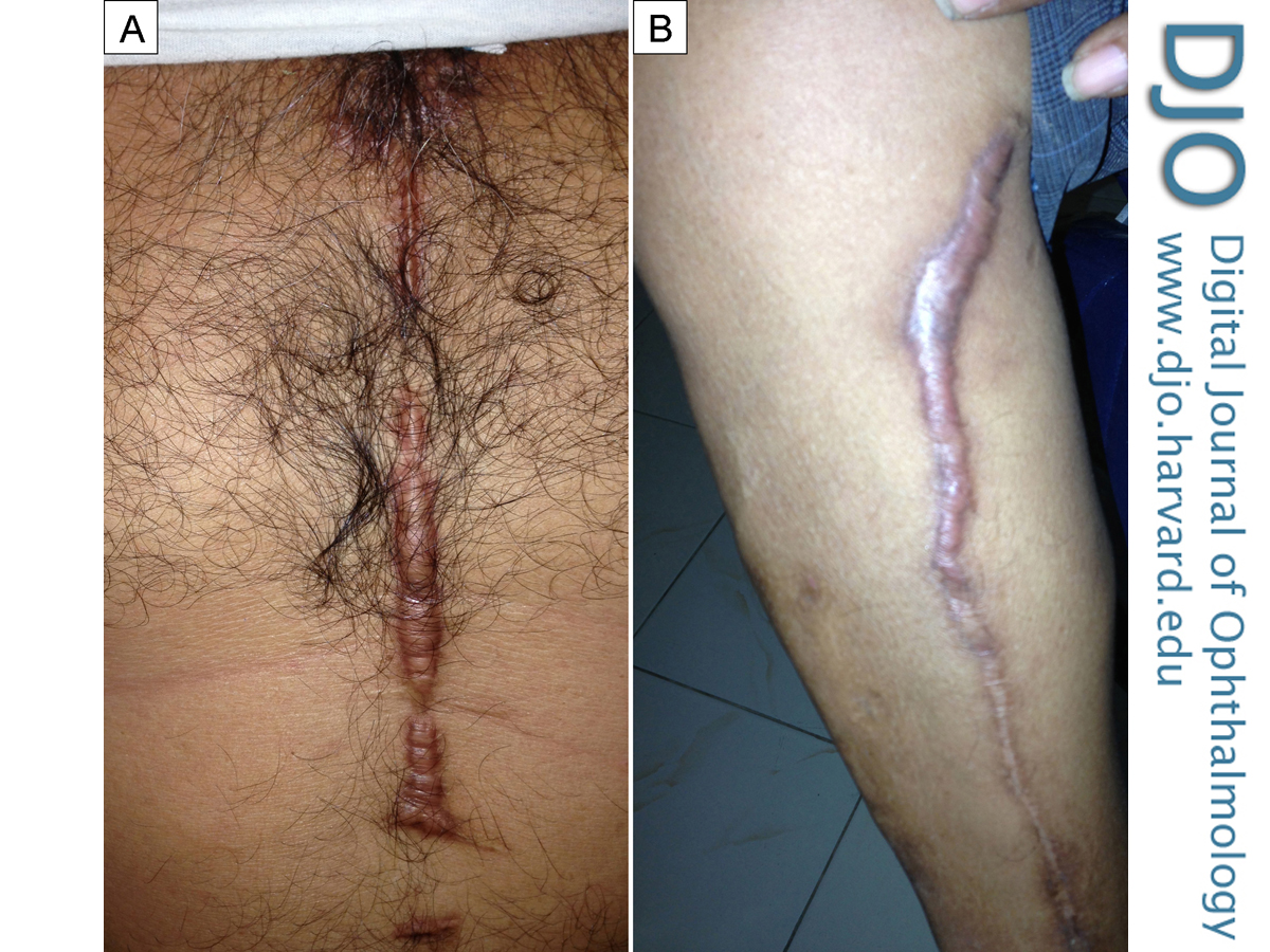
There was no history of uveitis, psudoexfoliation, or high myopia. Systemic examination revealed a scar of cardiac surgery on the chest with keloid formation at some places (Figure2A) His left leg showed a hypertrophic scar along the course of saphenous vein (Figure 2B). The patient was not diabetic.
Given the findings, anterior capsular peeling was undertaken. Proparacaine hydrochloride 0.5% (Paracain, Sunways Pvt Ltd, Mumbai, India) local anesthetic drops were instilled 3 times at 5-minute intervals; 0.5 mL of lidocaine hydrochloride 2% (Lox, Neon 41 Laboratories Ltd, Mumbai, India) was injected subconjunctivally. Video 1 shows the procedure.
A side port incision was made at the 12 o’clock position. Sodium hyaluronate 1.4% (Cohevisc, Appasamy Ocular Devices, Puducherry, India) was injected to form the anterior chamber. A temporal clear corneal incision was opened with the aid of an iris repositor. Passing the iris repositor and injecting hydroxypropyl methylcellulose 2% (Appavisc, Appasamy Ocular Devices) at the same time achieved separation of the capsular fibrosis from the IOL. The fibrotic band was cut radially at the 6 o’clock position using fine-pointed angled Vannas scissors. Utrata forceps were used to peel off the anterior capsular fibrosis, and the IOL was rotated in the bag with the dialer to unfold the haptic. Viscoelastic removal was performed from the anterior chamber. The side port and main incision were stromally hydrated.
Postoperatively, the patient was treated with moxifloxacin 0.3% and prednisolone acetate 1% eyedrops, tapered over 4 weeks, and he regained visual acuity of 20/20, which remained stable at 6 months’ follow-up. The postoperative autorefractometion and retinoscopy showed plano spherical with −0.25 × 180.
IOL positioning is crucial for postoperative visual gain. Vision may be impaired in patients with ACC because of the complete occlusion of the capsulorhexis opening, IOL decentration, or IOL tilting. Nd:YAG laser anterior capsulotomy has been proposed as a treatment option;(7) however, capsular debris loosened by this method may enhance the risk for inflammation and recurrences.(8) Moreover, delayed dislocation of the IOL in the ciliary sulcus has been reported after Nd:YAG laser capsulotomy in cases of ACC caused by extension of the radial relieving incision to the periphery.(9) Hayashi et al have shown use of Nd:YAG laser for radial anterior capsule relaxing incisions helps in preventing corneal endothelial damage more than the circular pattern.(10) Koizumi et al advocated the surgical peeling of the fibrous membrane in cases of thick ACC.(11) Panagopoulos et al used long, straight, and pointed scissors to cut the fibrotic membrane.(12)
Thick fibrosis of the anterior capsule with induced ammetropia caused by a tilt in the IOL were the indicators for surgical intervention in our case. The patient’s visual acuity was stable at 20/20, and no recurrence of the fibrotic membrane was noted at the 6 months’ follow-up.
Ocular risk factors suggested for the development of ACC include pseudoexfoliation syndrome, uveitis, high myopia, retinitis pigmentosa, and zonular weakness. A small capsulorhexis size, failure to polish the inner surface of the anterior capsular margin to clean the lens epithelial cells, and the IOL design and material are some of the modifiable intraoperative risk factors in the development of anterior capsular fibrosis (ACF).(12) Systemic risk factors implicated in the development of ACC include diabetes mellitus and myotonic dystrophy. None of the risk factors mentioned were present in our case.
The pathogenic mechanism responsible for the development of ACC is unknown. Reyntjens et al proposed the presence of alpha-smooth muscle actin in the metaplastic lens epithelial cells (LECs) that lead to ACC.(13) Ultrastructurally, the LECs exhibit specific microfilaments that are morphological features of myofibroblasts.(14,15) Kurosaka et al demonstrated an abundance of alpha-smooth muscle actin-positive LECs in the fibrous membrane of ACF.(16) Ehrlich et al demonstrated similar types of alpha-smooth muscle actin expressing myofibroblasts in a hypertrophic scar.(17) The histopathological features of ACF and hypertrophic scars have identical features. A search of the literature retrieved no results concerning a correlation between anterior or posterior capsular fibrosis responses in patients with a tendency for hypertrophic scarring. Our patient had undergone coronary artery bypass surgery and had hypertrophic scars on the chest and leg. The graft for the bypass surgery was taken from his left leg, which also showed a hypertrophic scar.
ACC has been noted in all types of IOL material and haptic designs, suggesting that the material itself or the configuration of the haptic does not play a role in the development of capsular contraction.(18) Postoperative inflammatory responses have been implicated in the development of capsular contraction.(19) No postoperative inflammation was observed in our case on follow-up. Studies have proposed the disruption of the blood-aqueous barrier, with an increase in blood-derived cytokines in the aqueous that results in the activation of LECs with fibrosis and in the contracture of the anterior capsule.(4,6) We hypothesize that an exaggerated fibrotic response from the anterior capsular margin in this patient could have caused the early ACC. Folding of the stiff haptic of the IOL on the optic indicated marked capsular contraction.
ACC has been reported in hydrophilic lenses with a stiff haptic (Rayner design),(11) plate haptic,(20) and C-loop haptic.(21) The pliability and softening of the plate and C-loop haptic result in buckling up over the optic, easily leading to the decentration of the IOL. Our patient had a stiff Rayner type haptic. To our knowledge, folding of this type of haptic over the optic has not been reported in the literature previously.
Our report highlights the possibility that severe and early ACC occluding the capsulorhexis opening can develop in a patient with a tendency for hypertrophic scar formation. Surgical intervention was necessary to correct IOL tilt-induced astigmatism for optimum visual gain. Further study on the correlation between anterior lens capsular contraction in patients with a tendency toward hypertrophic scarring is warranted, because both hypertrophic scars and ACC are not rare.
Literature Search
Pubmed, MEDLINE, Copernicus, Google Scholar, OpenJGate, EBSCO Publishing’s Electronic Databases, Hinari, African Index Medicus, China National Knowledge Infrastructure, Exlibris – Primo Central, Infotrieve, Journal Guide, National Science Library, ProQuest, and TdNet were all searched, without language restriction, on December 19, 2013, using the following terms: anterior capsular fibrosis, intraocular lens decentration, anterior capsular contraction AND intraocular lens material, and hypertrophic scar.
1. Masket S. Postoperative complications of capsulorhexis. J Cataract Refractive Surg 1993;19:721-4.
2. Hayashi H, Hayashi K, Nakao F, Hayashi F. Anterior capsular contraction and intraocular lens dislocation in eyes with pseudoexfoliation syndrome. Br J Ophthalmol 1998;82:1429-32.
3. Lu ?ke C , Dietlein TS, Jacobi PC, Konen W, Krieglstein GK. Massive anterior capsule shrinkage after plate-haptic silicone lens implantation in uveitis. J Cataract Refract Surg 2001;27:333-6.
4. Hayashi K, Hayashi H, Matuo K, Nakao F, Hayashi F. Anterior capsular contraction and intraocular lens dislocation after implant surgery in eyes with retinitis pigmentosa. Ophthalmology 1998;105:1239-43.
5. Hayashi H, Hayashi K, Nakao F, Hayashi F. Area reduction in the anterior capsular opening in eyes of diabetes mellitus patient. J Cataract Refractive Surg 1998;24:1105-10.
6. Rosa N, Lanza M, De Bernardo M, Borrelli M, Politano L. Anterior capsule phimosis and capsular block syndrome in a patient with Steinert myotonic dystrophy: a case report. Cases J 2009;2:92-8.
7. Chawla JS, Shaikh MH. Neodymium:YAG laser parabolic anterior capsulotomy in extreme capsule contraction syndrome. J Cataract Refract Surg 1999;25:1415–17.
8. Panagopoulos A, Chalioulias K, Kirkby GR. A new approach in the surgical management of anterior capsular phimosis syndrome. Ophthalmic Res 2009;42:221-3.
9. Tuft SJ, Talks SJ. Delayed dislocation of foldable plate-haptic silicone lenses after Nd:YAG laser anterior capsulotomy. Am J Ophthalmol 1998;126:586-8.
10. Hayashi K, Yoshida M, Hirata A, Hayashi H. Anterior capsule relaxing incisions with neodymium:YAG laser for patients at high-risk for anterior capsule contraction. J Cataract Refract Surg 2011;37:97-103.
11. Koizumi K, Watanabe A, Koizumi N, Kinoshita S. Peeling the fibrous membrane from the anterior capsule for capsulorhexis contraction after phacoemulsification in aphakic patients. J Cataract Refract Surg 2002; 28:1728-32.
12. Malik A, Gupta N, Sood S.Capsular contraction syndrome following insertion of hydrophilic acrylic lens. Int Ophthalmol 2011;31:121-3.
13. Reyntjens B, Tassignon M-J, Marck EV. Capsular peeling in anterior capsule contraction syndrome: surgical approach and histopathological aspects. J Cataract Refract Surg 2004;30:908-12.
14. McDonnell PJ, Zarbin MA, Green WR. Posterior capsule opacification in pseudophakic eyes. Ophthalmology 1983;90:1548-53.
15. Cobo LM, Ohsawa E, Chandler D, Arguello R, George G. Pathogenesis of capsular opacification after extracapsular cataract extraction; an animal model. Ophthalmology 1984;91:857-63.
16. Kurosaka D, Ando I, Kato K, Oshima T, Kurosaka H, Yoshino M, et al. Fibrous membrane formation at the capsular margin in capsule contraction syndrome. J Cataract Refract Surg 1999;25:930-5.
17. Ehrlich HP, Desmoulière A, Diegelmann RF, et al. Morphological and immunochemical differences between keloid and hypertrophic scar. Am J Pathol 1994;145:105-13.
18. Martínez Toldos JJ, Artola Roig A, Chipont Benabent E. Total anterior capsule closure after silicone intraocular lens implantation. J Cataract Refract Surg 1996;22:269-71.
19. Vock L, Georgopoulos M, Neumayer T, Buehl W, Findl O. Effect of the hydrophilicity of acrylic intraocular lens material and haptic angulation on anterior capsule opacification. Br J Ophthalmol 91:476-80.
20. Lam HH, Visvaraja S. Spontaneous dislocation of intraocular lens as a late complication of uncomplicated cataract surgery: a case series. Clin Exp Optom 2012;95:99-102.
21. van der Linden JW, van der Meulen IJ, Mourits MP, Lapid-Gortzak R. In-the-bag decentration of a hydrophilic radially asymmetric multifocal intraocular lens secondary to capsule contraction. J Cataract Refract Surg 2013;39:642-4.
 Welcome, please sign in