|
|
 |
|
|
|
|
|
Cilium incarnatum externum
Digital Journal of Ophthalmology
2017
Volume 23, Number 1
February 13, 2017
DOI: 10.5693/djo.01.2014.02.003
|
Printer Friendly
Download PDF |
|
|
 Taru Dewan, MS, FRCSEd
 | Department of Ophthalmology, PGIMER & RML Hospital, New Delhi, India Chetna Sharma, MBBS | Department of Ophthalmology, PGIMER RML Hospital, New Delhi, India Harshika Chawla, MBBS | Department of Ophthalmology, PGIMER RML Hospital, New Delhi, India Renu Singhania, MBBS | Department of Ophthalmology, PGIMER RML Hospital, New Delhi, India Ananya Chatterjee, MBBS | Department of Ophthalmology, PGIMER RML Hospital, New Delhi, India
|
|
|
| Abstract | | A 25-year-old woman presented with a history of a cosmetically bothersome bulge over her left eyelid. She was diagnosed with cilium incarnatum externum. The subcutaneous cilium was surgically removed and examined. | | | Introduction | Cilium incarnatum externum, a benign lid condition, in which cilium grows subcutaneously under the lid skin, is both rare and infrequently reported.(1) This condition responds to surgical access and epilation, thereby removing the cosmetic blemish.
| | | Case Report | A 25-year-old presented at PGIMER & RML Hospital, New Delhi, with a chief complaint of a localized bulge over the left upper eyelid. There were no visual complaints, pain, or redness of the eyelid skin, but it was a cause of cosmetic concern. There was no history of recurrent rubbing of the eyes, eyelid lesion or trauma preceding the patient’s noticing this bulge.
On examination, a localized linear swelling of 3.5 mm was present just above the upper eyelid margin. A misdirected eyelash was seen running obliquely underneath the skin from the lid margin of the left upper eyelid, causing the bulge (Figure 1A). The rest of the eyelashes were of normal position and direction. Visual acuity was normal; there were no other symptoms and no other abnormal findings. A diagnosis of cilium incarnatum externum was made.
Surgical removal of the eyelash was planned. A small opening was made in the overlying skin using a lance-tip blade, and the cilium was removed using a Sinsky dialer. The cilium was 3 mm in length, thinner and smaller than the rest. It was heavily pigmented and curled on itself (Figure 1B). It ran in the subcutaneous space throughout its entire length. The entire follicle was misdirected, leading to a vertically upward course of the lash. It was epilated using forceps. The skin stab did not require suturing. Topical ciprofloxacin ointment was applied 3 times daily for 5 days.
| |
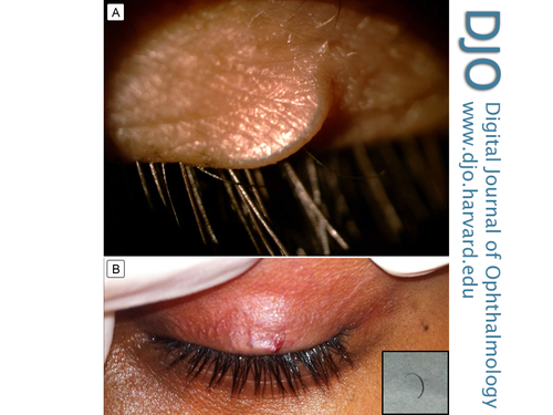
Figure 1
A, Subcutaneous elevation caused by cilium incarnatum externum. B, Exposed aberrant eyelash thinner and smaller than normal lashes, with epilated eye lash (inset).
|
|
| Discussion | Misdirected, ingrowing cilia can either be present subconjunctivally (cilium incarnatum internum) or subcutaneously (cilium incarnatum externum).(1) Cilium incarnatum externum can be a hereditary or acquired condition. The latter condition is thought to be a result of local pathology causing the misdirection of the lash or entire follicle.(2-4) In the hereditary variety, two patterns may be seen.(1) An eyelash after coming out obliquely from the follicle, again pierces the epithelium of the skin and grows underneath it. Sometimes, though, it grows beneath the epidermis throughout its whole course.
A review of literature revealed very few isolated reports. The reported cases were largely asymptomatic and presented either due to a cosmetic concern or mistaking the lesion to be a lid nodule or a blackhead.(3,5) The only symptom that has been described is a needlelike sensation when the area is rubbed.(6) Most reported cases occurred in young female subjects.
Previous cases have described the morphology of the abnormal eyelash as similar to other normal eyelashes, except visible heavy pigmentation and increased thickness in 1 case.(5,7) In our case, the eyelash, though pigmented, was smaller, thinner, and curved as compared to other lashes. The last case reported was in 1976.(8) No further cases have been reported. Awareness of such a condition can prompt simple corrective measures by ophthalmologists rather than mishandling by the overzealous patient or cosmetologist.
Literature Search
The authors performed an English-language search of PubMed, HubMed, and Google Scholar using the following terms: cilium incarnatum externum and ingrowing eyelash. Cross references were checked for related articles. | | | References | 1. Duke-Elder S, ed. System of Ophthalmology. Vol. 13. St Louis: Mosby; 1974:386.
2. Agarwala HS. Cilium inversum. Am J Ophthalmol 1963;55:648-9.
3. Bloch FJ. Minor anomalies in position of the eyelashes. Arch Ophthalmol 1947;37:772-4.
4. Herzog H. Pathologie der Cilien, Z Augenheilkd 1904;12:256.
5. Makrocki F. Ein Fall von pervers gewachsenen subcutanen Cilien. Zentralbl prakt Augenh 1883;7:129.
6. Sen DK, Mohan H, Gupta DK. Cilium inversum. Br J Ophthalmol 1969;53:207.
7. Cherno F. Über die unter dem Namen Blepharitis ciliaris bekannten Erkrankungen des Lidrandes, Z Augenheilkd 1907;18:1.
8. Belfort R, Ostler HB. Cilia incarnata. Br J Ophthalmol 1976;60:594-6.
| |
|
|
|
|
|
|
 Welcome, please sign in


