|
|
 |
|
|
|
|
|
Clostridium septicum endophalmitis associated with colon adenocarcinoma
Digital Journal of Ophthalmology
2014
Volume 20, Number 3
September 7, 2014
DOI: 10.5693/djo.02.2014.01.001
|
Printer Friendly
Download PDF |
|
|

Jacob L. Eisenrich, BS | Department of Ophthalmology, University of Texas Health Science Center, San Antonio, Texas Angela M. Herro, MD | Department of Ophthalmology, University of Texas Health Science Center, San Antonio, Texas Mason Schmutz, MD | Department of Ophthalmology, University of Texas Health Science Center, San Antonio, Texas Kundandeep S. Nagi, MD | Department of Ophthalmology, University of Texas Health Science Center, San Antonio, Texas
|
|
|
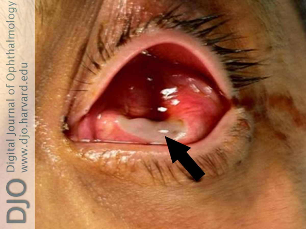
| Abstract | | Clostridium endophthalmitis is a rare but serious infection. Most cases are associated with exogenous causes, such as inoculation secondary to trauma. However, endogenous cases do occur. Clostridium septicum is the primary clostridial pathogen associated with endogenous endophthalmitis. These infections have been associated with malignancy, particularly colonic and hematologic. We present a case of endogenous Clostridium septicum endophthalmitis associated with adenocarcinoma of the colon. This case highlights the value of prompt recognition of endogenous endophthalmitis and concurrent systemic evaluation. | | | Introduction | | Clostridium endophthalmitis is a rare but serious infection.(1) It usually occurs following a penetrating injury of the globe and is most often caused by Clostridium perfringens.(2,3) Endogenous causes also pose a serious threat to vision and can herald other serious illnesses. Endogenous endophalmitis caused by clostridial species appear to have a predilection for enteric sources.(4) Clostridium septicum is the primary pathogen associated with endogenous clostridial endophthalmitis. Koransky et al. report that 71% of Clostridium septicum bacteremia cases are associated with malignancies.(5) Only 7 cases have been reported in the English language literature, the first in 1985.(3,6) | | | Case Report | A 56-year-old man with poorly controlled diabetes, hypertension, asthma, and hepatitis B presented to the Emergency Department at University of Texas Health Science Center, San Antonio, with 1 week of left-eye pain, bleeding, and discharge of viscous, dark fluid. Examination of the left eye revealed a deflated globe with conjunctival injection, mucopurulent discharge, and an opacified, indented cornea with a central perforation expressing a brown liquid (Figure 1). Visual acuity testing revealed no light perception and examination of the fundus was not possible. Examination of the right eye was normal.
On further questioning, the patient indicated that he had self-treated a recent flare-up of asthma with oral prednisone and that he had experienced diffuse, dull, right upper quadrant abdominal pain. Additionally, approximately 1 week prior to presenting, he was treated at an outpatient clinic with fortified topical vancomycin and tobramycin eyedrops for a corneal infiltrate in the left eye. He subsequently developed a purulent discharge and was treated with intramuscular ceftriaxone for suspected gonorrheal keratoconjunctivitis. His condition continued to decline in the following days and intravitreal vancomycin and ceftazidime were administered approximately 2 days prior to his presentation at the Emergency Department.
The patient immediately underwent primary repair of the perforation. Intraoperatively the cornea was found to be completely necrotic, with a central perforation. Due to lack of viable tissue the decision was made to remove the cornea. This allowed visualization of the posterior pole; however the contents were unidentifiable. The lens was identified in the posterior pole and removed. Liquefied vitreous was sampled and sent for evaluation. The eye was irrigated with antibiotic and closed, limbus to limbus with 8-0 nylon. Tenons and conjunctiva were subsequently closed with 8-0 polyglactin 910 suture. Moxifloxacin drops and intravenous vancomycin and piperacillin-tazobactam were initiated. Histopathological analysis revealed the presence of moderate Gram-positive and rare Gram-negative rods. Subsequent testing led to the discovery of iron-deficiency anemia and guaiac-positive stool. A computed tomography (CT) scan of the abdomen revealed a localized abnormality near the splenic flexure of the colon and a normal aorta. The patient was scheduled for a colonoscopy following discharge.
One week after initial repair, he was found to have a small segment of wound dehiscence and uveal prolapse with a mild, purulent discharge. At this point, the left eye was eviscerated. Tissue cultures from the initial repair and the uveal sample grew Clostridium septicum.
The colonoscopy revealed adenocarcinoma, and the patient underwent partial colon resection followed by 6 months of chemotherapy. At last follow-up, 6 months after completion of chemotherapy, there was no evidence of disease. | |

Figure 1
The patient’s left eye at presentation to the emergency department showing a deflated eye. The cornea is opacified and perforated (black arrow).
|
|
| Discussion | Signs and symptoms of endophthalmitis from the histotoxic species (C. perfringens, C. septicum) include chemosis, swelling of the eyelids, hypopyon, ring-abscess of the cornea, coffee-colored discharge, gas bubbles in the anterior chamber, and loss of the fundus reflex.(1,7,8) Our patient exhibited a profound corneal abscess, loss of the fundus reflex, and coffee-colored discharge.
A seminal study of Clostrium septicum bacteremia showed that 71% of the cases were associated with malignancies, equally divided between solid tumors and hematologic malignancies; of those with solid tumors, 67% had colon cancer.(5) The distal ileum or cecum has been identified as the likely location of entry into the blood stream in these cases.(5) Bacteremia without a clear portal of entry may indicate the presence of an occult tumor.(1) Consistent with other studies, our patient had diabetes mellitus type II, hypertension, and may have been immunosuppressed as a consequence of the prednisone he had been taking for asthma.(2) His ocular infection ultimately led to the discovery of adenocarcinoma of the colon, a potentially fatal, systemic illness.
Patients with clostridial infections may deteriorate quickly.(9) Our patient’s tumor was caught relatively early and removed before lymphatic spread.
Literature Search
PubMed was searched for English-language articles on December 15, 2012, using the following terms: clostridium endophthalmitis, clostridial bacteremia, endogenous clostridium, clostridium septicum, and bacterial endophthalmitis. Sources in retrieved articles were cross-referenced. | | | References | 1. Fejes I, Dégi R, Végh M. Clostridium septicum gas gangrene in the orbit: a case report. Infection 2013;41:276-70.
2. Cannistra AJ, Albert DM, Frambach DA, Dreher RJ, Roberts L. Sudden visual loss associated with clostridial bacteraemia. Br J Ophthalmology 1988;72:380-5.
3. Bradley JC, Kimbrough RC, Vidal AM, McCartney DL, Sarria JC. Endophthalmitis caused by non-perfringens Clostridium species. Scand J Infect Dis 2004;36:689-92.
4. Nangia V, Hutchinson C. Metastatic endophthalmitis caused by Clostridium perfringens. Br J Ophthalmol 1992;76:252-3.
5. Koransky JR, Stargel MD, Dowell VR. Clostridium septicum bacteremia: its clinical significance. Am J Med 1979;66:63-6.
6. Lindland A, Slagsvold JE. Binocular endogenous Clostridium septicum endophthalmitis. Acta Ophthalmol Scand 2007;85:232-4.
7. Leavelle RB. Gas gangrene panophthalmitis; review of the literature; report of new cases. AMA Arch Ophthalmol 1955;53:634-42.
8. Lauer AK, Riley K, Wentzien J, Marsal SW. Acute painful vision loss and acute abdomen: a case of endogenous Clostridium perfringens endophthalmitis. Can J Ophthalmol 2005;40:208-10.
9. Shwe-Tin A, Ung T, Madhavan C, Yasen T. A case of endogenous Clostridium perfringens endophthalmitis in an intravenous drug abuser. Eye (Lond) 2007;21:1427-8.
| |
|
|
|
|
|
|
 Welcome, please sign in