|
|
 |
|
|
|
|
|
A rare and unusual retinal arterioarterial communication in a prepapillary vascular loop
Digital Journal of Ophthalmology
2014
Volume 20, Number 1
February 10, 2014
DOI: 10.5693/djo.02.2014.01.002
|
Printer Friendly
Download PDF |
|
|
|
|
|
|
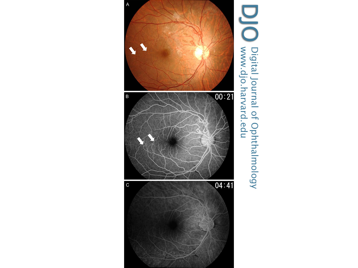
| Abstract | | Fundus examination of a 40-year-old man presenting with blurred vision in his right eye revealed mild vitreous hemorrhage, a prepapillary vascular loop, and a peripapillary epiretinal membrane. Additionally, a superotemporal branch retinal artery communicated directly with an inferotemporal branch retinal artery in the temporal fovea at 2 locations. Fluorescein angiography confirmed an extremely rare arterioarterial communication. Two weeks later the vitreous hemorrhage and small intraretinal hemorrhage had disappeared, and the visual acuity in the right eye improved. | | | Introduction | | Prepapillary vascular loop formations are uncommon congenital vascular malformations, the majority of which are of arterial origin.(1) They are usually detected incidentally on routine fundus examination but have been associated with retinal vascular occlusive disease and recurrent vitreous bleeding. The incidence of prepapillary vascular loops ranges from approximately 1 in 2000 to 1 in 9000 patients; 9%-17% of these cases are bilateral.(2) Congenital arteriovenous communications or malformations are rare retinal vascular anomalies;(3) retinal arterioarterial communications are extremely rare. Prepapillary vascular loops associated with arterioarterial communication have been reported rarely.(4,5) This report describes a case of this rare combination. | | | Case Report | | A 40-year-old man was referred to Jichi Medical University Hospital with blurred vision in his right eye of one day’s duration. His personal history and family history were noncontributory; systemic evaluation was unremarkable. On ophthalmological examination, visual acuity was 20/40 in the right eye and 20/15 in the left eye. Anterior segment examination was normal in both eyes. Intraocular pressure was normal in each eye. Fundus examination of the right eye showed mild vitreous hemorrhage, a prepapillary vascular loop, a peripapillary epiretinal membrane, and a small intraretinal hemorrhage (Figure 1A). Additionally, a superotemporal branch retinal artery communicated directly with an inferotemporal branch retinal artery in the temporal fovea at 2 locations. Fluorescein angiography confirmed an unusual arterioarterial communication (Figure 1B). In the late phase, there was no dye leakage and no staining at the peripapillary vascular loop, the arterioarterial communication, and the other retinal vessels (Figure 1C). Optical coherence tomography imaging was unavailable. Two weeks after the initial visit, the vitreous hemorrhage and small intraretinal hemorrhage had disappeared; visual acuity in the right eye improved to 20/15. | |
Figure 1
Fundus photograph (A) and fluorescein angiogram (B) of the right eye (21 seconds after injection) showing a prepapillary vascular loop and the communication of superotemporal and inferotemporal branch retinal arteries (arrows). A late phase of a fluorescein angiogram (C) of the right eye (281 seconds after injection) showing no dye leakage and no staining.
|
|
| Discussion | Various types of congenital and acquired retinal vascular communications have been described.(1-3,6) Acquired retinal arteriovenous communications are recognized manifestations of ischemic and occlusive diseases. Retinal arterioarterial communications are rare; the coexistence of retinal arterioarterial communication and prepapillary vascular loops is extremely rare.(4,5,7,8) Nishimura et al described a 27-year-old man with arteroarterial communication at the peripapillary region.(4) Tamura et al described a 25-year-old man with arterioarterial communication; however, the patient had a history of branch retinal artery occlusion.(5) Two cases of arterioarterial communications with angioid streaks have been reported,(7,8) both showing vascular loop formation. This is the first reported case of a patient with arterioarterial communication, located at the posterior pole of the eye, and a prepapillary vascular loop. In addition, a peripapillary epiretinal membrane was detected. I speculate that vitreous hemorrhage may have occurred from the superficial retinal capillaries at the vascular loop during an acute posterior vitreous detachment.
Retinal racemose hemangiomas are congenital retinal vascular malformations with anomalous arteriovenous communications and shunts that result from misdifferentiation of the embryological vasculature during vasculogenesis.(6) Archer et al noted that although the pathogenesis of these isolated arteriovenous malformations is unknown,(3) they probably represent a local defect in the maturation of the primitive retinal mesenchymal cells, which originate from the hyaloid vascular system and differentiate into solid cords of endothelial cells. Vascular abnormalities in rasemose hemangioma consist of arteriovenous malformations. Arterioarterial communication should be distinguished from arteriovenous communication; however, some vascular loop formation is congenital. Detailed study of additional cases is necessary to confirm the hypothesized relation between the vascular loop, rasemose hemangioma, and retinal arterioarterial communication.
Literature Search
PubMed was searched on September 20, 2014, for English-language articles (1975- present) using the following combinations of terms: prepapillary vascular loop; arterioarterial communication AND retina. In addition, the author also searched Ichushi Service on September 20, 2014, for Japanese-language article (1983-present) using the following search terms: prepapillary vascular loop, communication of the retinal vessel. | | | References | 1. Degenhart W, Brown GC, Augsburger JJ, Magargal L. Prepapillary vascular loops. Ophthalmology 1981;88:1126-31.
2. Romano PE. Prepapillary vascular loops. Clin Exp Ophthalmol 2001;29:90-1.
3. Archer DB, Deutman A, Ernest JT, Krill AE. Arteriovenous communications of the retina. Am J Ophthalmol 1973;75:224-41.
4. Nishimura T, Uyama M. Retinal vascular loop formation on the optic disc. Jap J Clin Ophthalmol 1982;36:1371-5.
5. Tamura M, Atsumi O, Yoshimoto H. Retinal vascular loop formation on the optic disc: retrospective study of eleven cases. Folia Ophthalmol Jap 1991;42:304-10.
6. Schmidt D, Pache M, Schumacher M. The congenital unilateral retinocephalic vascular malformation syndrome (Bonnet-Dechaume-Blanc syndrome or Wyburn-Mason syndrome): review of the literature. Surv Ophthalmol 2008;53:227-49.
7. Khairallah M. Congenital communication of cilioretinal and retinal arteries associated with angioid streaks. Arch Ophthalmol 1997;115:1328-9.
8. Secrétan M, Zografos L, Guggisberg D, Piguet B. Chorioretinal vascular abnormalities associated with angioid streaks and pseudoxanthoma elasticum. Arch Ophthalmol 1998;116:1333-6.
| |
|
|
|
|
|
|
 Welcome, please sign in